Minhaj Shaikh, Sarbesh Tiwari, Taruna Yadav, Pawan K Garg, Pushpinder S Khera
{"title":"Leukoencephalopathy with Calcifications and Cysts in a Child with Progressive Hemiparesis-A Case Report.","authors":"Minhaj Shaikh, Sarbesh Tiwari, Taruna Yadav, Pawan K Garg, Pushpinder S Khera","doi":"10.4103/jpn.JPN_113_20","DOIUrl":null,"url":null,"abstract":"<p><p>With the advent of modern neuroimaging, the imaging features of various leukoencephalopathies have been unraveled in the past two decades. Leukoencephalopathy with calcifications and cysts (LCC) is one such rare autosomal recessive disorder with marked clinical heterogeneity and a striking but characteristic imaging appearance-diffuse white matter changes, intraparenchymal cysts, and calcifications. The calcifications in LCC are characteristically nodular, dense, bulky, and predominantly located in gray nuclei of the central brain (basal ganglia, thalami) and cerebellum (dentate nuclei). We describe a case of a 9-year-old boy with progressive left hemiparesis and seizures, which on imaging showed characteristic features of LCC. We further review the neuroimaging features of LCC and its differential diagnoses.</p>","PeriodicalId":46746,"journal":{"name":"Journal of Pediatric Neurosciences","volume":"16 4","pages":"277-280"},"PeriodicalIF":0.2000,"publicationDate":"2021-10-01","publicationTypes":"Journal Article","fieldsOfStudy":null,"isOpenAccess":false,"openAccessPdf":"https://www.ncbi.nlm.nih.gov/pmc/articles/PMC9757512/pdf/","citationCount":"0","resultStr":null,"platform":"Semanticscholar","paperid":null,"PeriodicalName":"Journal of Pediatric Neurosciences","FirstCategoryId":"1085","ListUrlMain":"https://doi.org/10.4103/jpn.JPN_113_20","RegionNum":0,"RegionCategory":null,"ArticlePicture":[],"TitleCN":null,"AbstractTextCN":null,"PMCID":null,"EPubDate":"","PubModel":"","JCR":"Q3","JCRName":"Medicine","Score":null,"Total":0}
引用次数: 0
Abstract
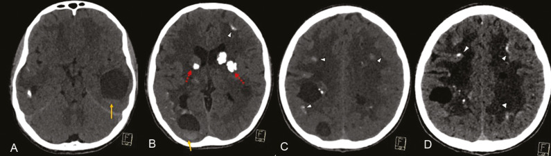
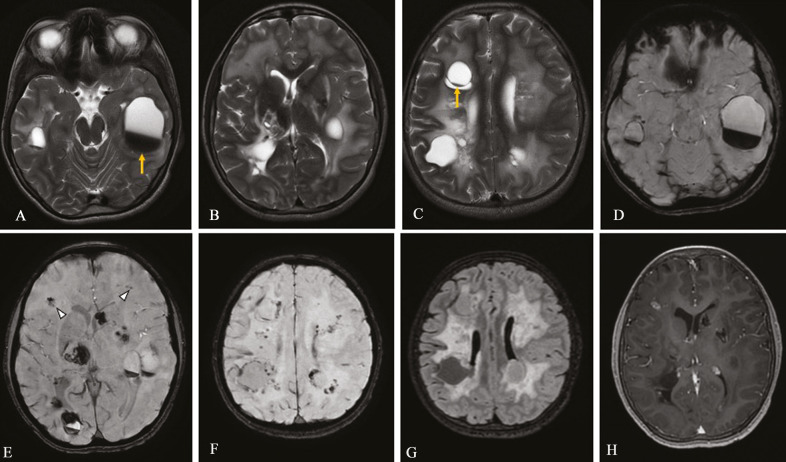
With the advent of modern neuroimaging, the imaging features of various leukoencephalopathies have been unraveled in the past two decades. Leukoencephalopathy with calcifications and cysts (LCC) is one such rare autosomal recessive disorder with marked clinical heterogeneity and a striking but characteristic imaging appearance-diffuse white matter changes, intraparenchymal cysts, and calcifications. The calcifications in LCC are characteristically nodular, dense, bulky, and predominantly located in gray nuclei of the central brain (basal ganglia, thalami) and cerebellum (dentate nuclei). We describe a case of a 9-year-old boy with progressive left hemiparesis and seizures, which on imaging showed characteristic features of LCC. We further review the neuroimaging features of LCC and its differential diagnoses.
期刊介绍:
Journal of Pediatric Neurosciences-JPN (ISSN 1817-1745) is official publication of the Indian Society for Pediatric Neurosurgery. The journal is published semiannually. Bibliographic listings: The journal is indexed with Caspur, DOAJ, EBSCO Publishing’s Electronic Databases, Excerpta Medica / EMBASE, Expanded Academic ASAP, Genamics JournalSeek, Google Scholar, Health & Wellness Research Center, Health Reference Center Academic, Hinari, Index Copernicus, OpenJGate, Scimago Journal Ranking, SCOLOAR, SCOPUS, SIIC databases, Ulrich’s International Periodical Directory


 分享
分享
 求助内容:
求助内容: 应助结果提醒方式:
 扫码关注我们
