Zhiyan Fu, Evita B Henderson-Jackson, Barbara A Centeno, Gregory Y Lauwers, Mihaela Druta, Daniel A Anaya, Yukihiro Nakanishi
{"title":"A Case of Hepatic Malignant Solitary Fibrous Tumor: A Case Report and Review of the Literature.","authors":"Zhiyan Fu, Evita B Henderson-Jackson, Barbara A Centeno, Gregory Y Lauwers, Mihaela Druta, Daniel A Anaya, Yukihiro Nakanishi","doi":"10.1155/2023/2271690","DOIUrl":null,"url":null,"abstract":"<p><p>A 73-year-old man with a history of atrial myxoma and basal cell carcinoma presented with unexplained fever. Contrast-enhanced CT abdomen showed a large left hepatic lobe mass with early enhancement and delayed venous washout, concerning for hepatocellular carcinoma. Fine needle aspiration showed numerous spindle cells with malignant nuclear features, suggestive of malignant spindle cell neoplasm. The patient underwent left hepatectomy. The surgical specimen showed a well-circumscribe solid mass (14.6 × 13.0 × 10.0 cm) with necrosis. Histopathological examination revealed a proliferation of spindle tumor cells with characteristic staghorn-shaped blood vessels, frequent mitoses, and necrosis. The tumor cells showed strong and diffuse expression of CD34 and STAT6, confirming the diagnosis of malignant solitary fibrous tumor. Solitary fibrous tumor is a rare fibroblastic tumor characterized by a staghorn vasculature and NAB2-STAT6 gene rearrangement. Solitary fibrous tumor of the liver is a rare occurrence. Although most solitary fibrous tumors behave in a benign fashion, solitary fibrous tumors might act aggressively. This case is unique in that it demonstrates an excellent correlation between radiologic, macroscopic, and microscopic features which can contribute to the improvement of radiologic and pathologic diagnostic accuracy.</p>","PeriodicalId":45638,"journal":{"name":"Case Reports in Pathology","volume":"2023 ","pages":"2271690"},"PeriodicalIF":0.5000,"publicationDate":"2023-01-01","publicationTypes":"Journal Article","fieldsOfStudy":null,"isOpenAccess":false,"openAccessPdf":"https://www.ncbi.nlm.nih.gov/pmc/articles/PMC9935885/pdf/","citationCount":"0","resultStr":null,"platform":"Semanticscholar","paperid":null,"PeriodicalName":"Case Reports in Pathology","FirstCategoryId":"1085","ListUrlMain":"https://doi.org/10.1155/2023/2271690","RegionNum":0,"RegionCategory":null,"ArticlePicture":[],"TitleCN":null,"AbstractTextCN":null,"PMCID":null,"EPubDate":"","PubModel":"","JCR":"Q4","JCRName":"PATHOLOGY","Score":null,"Total":0}
引用次数: 0
Abstract
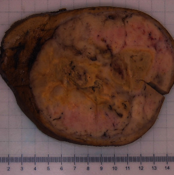
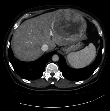
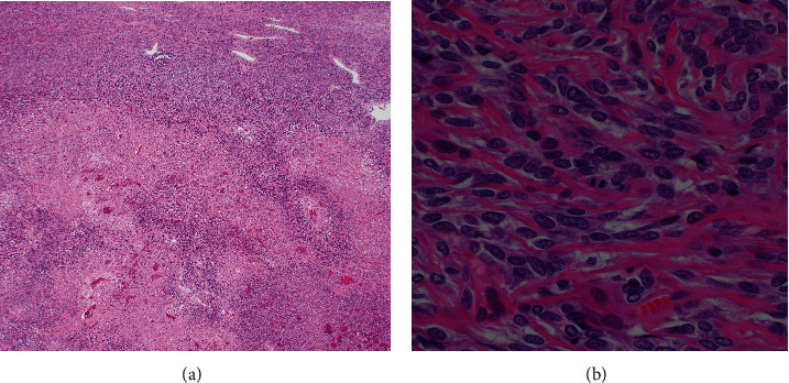
A 73-year-old man with a history of atrial myxoma and basal cell carcinoma presented with unexplained fever. Contrast-enhanced CT abdomen showed a large left hepatic lobe mass with early enhancement and delayed venous washout, concerning for hepatocellular carcinoma. Fine needle aspiration showed numerous spindle cells with malignant nuclear features, suggestive of malignant spindle cell neoplasm. The patient underwent left hepatectomy. The surgical specimen showed a well-circumscribe solid mass (14.6 × 13.0 × 10.0 cm) with necrosis. Histopathological examination revealed a proliferation of spindle tumor cells with characteristic staghorn-shaped blood vessels, frequent mitoses, and necrosis. The tumor cells showed strong and diffuse expression of CD34 and STAT6, confirming the diagnosis of malignant solitary fibrous tumor. Solitary fibrous tumor is a rare fibroblastic tumor characterized by a staghorn vasculature and NAB2-STAT6 gene rearrangement. Solitary fibrous tumor of the liver is a rare occurrence. Although most solitary fibrous tumors behave in a benign fashion, solitary fibrous tumors might act aggressively. This case is unique in that it demonstrates an excellent correlation between radiologic, macroscopic, and microscopic features which can contribute to the improvement of radiologic and pathologic diagnostic accuracy.


 分享
分享
 求助内容:
求助内容: 应助结果提醒方式:
 扫码关注我们
