Malgorzata Polacin, Tobias Hünermund, Oliver Müggler, Hatem Alkadhi, Sebastian Kozerke, Robert Manka
{"title":"Patient-Specific Cardiac Magnetic Resonance Feature Tracking Approach for Scar Detection in Concomitant Ischemic and Non-Ischemic Heart Disease.","authors":"Malgorzata Polacin, Tobias Hünermund, Oliver Müggler, Hatem Alkadhi, Sebastian Kozerke, Robert Manka","doi":"10.26502/fccm.92920297","DOIUrl":null,"url":null,"abstract":"<p><strong>Aim: </strong>This study investigated a patient-specific approach of using cardiac magnetic resonance (CMR) feature tracking for scar detection in a heterogenous patient group with chronic ischemic and non-ischemic heart disease.</p><p><strong>Methods: </strong>CMR exams of 89 patients with concomitant chronic ischemic and non-ischemic heart disease (IHD+) as well as 65 patients with ischemic scars only (IHD) were retrospectively evaluated. In all patients, global (GCS) and segmental circumferential strain (SCS) was derived from native cine images using a dedicated software (Segment CMR, Medviso). After calculation of patient-specific median GCS (GCS<sub>median</sub>), segmental values from GCS<sub>median</sub> percentage plots were correlated with corresponding myocardial segments in late gadolinium enhancement (LGE).</p><p><strong>Results: </strong>Overall GCS ranged between -3.5% to -19.8% and average GCS was lower in IHD+ than in IHD (p <0.05). In IHD, 19% of all myocardial segments were infarcted, in IHD+ 16.6%. Additionally, non-ischemic LGE was present in 6.7% of segments in IHD+. Correlation of GCS<sub>median</sub> percentage plots with corresponding LGE showed that presence of ischemic scar tissue in a myocardial segment was very likely below a cut-off of 39.5% GCS<sub>median</sub> (87.5% sensitivity, 86.3% specificity, AUC 0.907, 95% CI 0.875-0.938, p < 0.05).</p><p><strong>Conclusion: </strong>In patient-specific GCS<sub>median</sub> percentage plots calculated from native cine images, ischemic scar tissue can be suspected in myocardial segments below the threshold of 40% GCS<sub>median</sub> (sensitivity 88%, specificity 86%), even in a heterogenous patient cohort with ischemic and non-ischemic heart disease.</p>","PeriodicalId":72523,"journal":{"name":"Cardiology and cardiovascular medicine","volume":"6 6","pages":"542-549"},"PeriodicalIF":0.0000,"publicationDate":"2022-01-01","publicationTypes":"Journal Article","fieldsOfStudy":null,"isOpenAccess":false,"openAccessPdf":"https://www.ncbi.nlm.nih.gov/pmc/articles/PMC9937585/pdf/","citationCount":"0","resultStr":null,"platform":"Semanticscholar","paperid":null,"PeriodicalName":"Cardiology and cardiovascular medicine","FirstCategoryId":"1085","ListUrlMain":"https://doi.org/10.26502/fccm.92920297","RegionNum":0,"RegionCategory":null,"ArticlePicture":[],"TitleCN":null,"AbstractTextCN":null,"PMCID":null,"EPubDate":"","PubModel":"","JCR":"","JCRName":"","Score":null,"Total":0}
引用次数: 0
Abstract
Aim: This study investigated a patient-specific approach of using cardiac magnetic resonance (CMR) feature tracking for scar detection in a heterogenous patient group with chronic ischemic and non-ischemic heart disease.
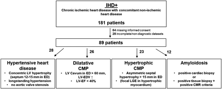
Methods: CMR exams of 89 patients with concomitant chronic ischemic and non-ischemic heart disease (IHD+) as well as 65 patients with ischemic scars only (IHD) were retrospectively evaluated. In all patients, global (GCS) and segmental circumferential strain (SCS) was derived from native cine images using a dedicated software (Segment CMR, Medviso). After calculation of patient-specific median GCS (GCSmedian), segmental values from GCSmedian percentage plots were correlated with corresponding myocardial segments in late gadolinium enhancement (LGE).
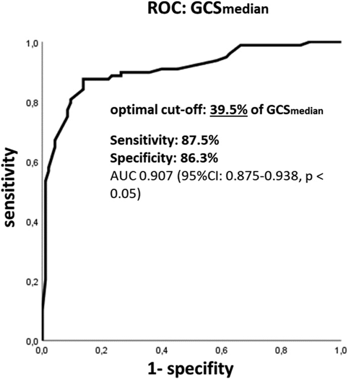
Results: Overall GCS ranged between -3.5% to -19.8% and average GCS was lower in IHD+ than in IHD (p <0.05). In IHD, 19% of all myocardial segments were infarcted, in IHD+ 16.6%. Additionally, non-ischemic LGE was present in 6.7% of segments in IHD+. Correlation of GCSmedian percentage plots with corresponding LGE showed that presence of ischemic scar tissue in a myocardial segment was very likely below a cut-off of 39.5% GCSmedian (87.5% sensitivity, 86.3% specificity, AUC 0.907, 95% CI 0.875-0.938, p < 0.05).
Conclusion: In patient-specific GCSmedian percentage plots calculated from native cine images, ischemic scar tissue can be suspected in myocardial segments below the threshold of 40% GCSmedian (sensitivity 88%, specificity 86%), even in a heterogenous patient cohort with ischemic and non-ischemic heart disease.


 分享
分享

 求助内容:
求助内容: 应助结果提醒方式:
 扫码关注我们
