{"title":"Epstein‑Barr virus‑associated lymphoepithelial carcinoma arising in the parotid gland: A case report and literature review.","authors":"Akinobu Kubota, Nobuyuki Bandoh, Takashi Goto, Ken-Ichi Matsumoto, Tomomi Yamaguchi-Ishochi, Yasutaka Kato, Hiroshi Nishihara, Hidehiro Takei","doi":"10.3892/mco.2023.2620","DOIUrl":null,"url":null,"abstract":"<p><p>A 60-year-old woman presented with a 3-year history of a slow-growing, painless mass in their left parotid gland. Ultrasonography revealed a well-circumscribed, lobulated, hypoechoic mass measuring 19x12x10 mm in the left parotid gland. Computed tomography revealed a well-circumscribed, solid mass with homogeneous enhancement. Fluorodeoxyglucose-positron emission tomography revealed uptake by the tumor but no uptake in other organs, including the nasopharynx. The patient underwent superficial parotidectomy with adequate safety margins and selective neck dissection followed by radiotherapy. No facial paralysis or recurrence of the tumor had been observed as of 20 months post-operation. Histologically, the tumor was composed of sheets of syncytial cancer cells with prominent nucleoli in a dense lymphoplasmacytic background. Epstein-Barr virus (EBV)-encoded RNA <i>in situ</i> hybridization was diffusely positive in the tumor cells. These findings indicated that the tumor was an EBV-associated lymphoepithelial carcinoma. Metastasis, especially from the nasopharynx, was excluded endoscopically and radiologically. Targeted next-generation sequencing of 160 cancer-related genes using the surgical specimen revealed no mutations, including known significant mutations detected in EBV-associated nasopharyngeal carcinoma.</p>","PeriodicalId":18737,"journal":{"name":"Molecular and clinical oncology","volume":"18 3","pages":"24"},"PeriodicalIF":1.4000,"publicationDate":"2023-03-01","publicationTypes":"Journal Article","fieldsOfStudy":null,"isOpenAccess":false,"openAccessPdf":"https://ftp.ncbi.nlm.nih.gov/pub/pmc/oa_pdf/ee/85/mco-18-03-02620.PMC9944707.pdf","citationCount":"1","resultStr":null,"platform":"Semanticscholar","paperid":null,"PeriodicalName":"Molecular and clinical oncology","FirstCategoryId":"1085","ListUrlMain":"https://doi.org/10.3892/mco.2023.2620","RegionNum":0,"RegionCategory":null,"ArticlePicture":[],"TitleCN":null,"AbstractTextCN":null,"PMCID":null,"EPubDate":"","PubModel":"","JCR":"Q4","JCRName":"ONCOLOGY","Score":null,"Total":0}
引用次数: 1
Abstract
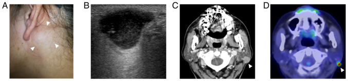
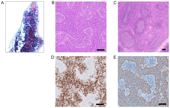
A 60-year-old woman presented with a 3-year history of a slow-growing, painless mass in their left parotid gland. Ultrasonography revealed a well-circumscribed, lobulated, hypoechoic mass measuring 19x12x10 mm in the left parotid gland. Computed tomography revealed a well-circumscribed, solid mass with homogeneous enhancement. Fluorodeoxyglucose-positron emission tomography revealed uptake by the tumor but no uptake in other organs, including the nasopharynx. The patient underwent superficial parotidectomy with adequate safety margins and selective neck dissection followed by radiotherapy. No facial paralysis or recurrence of the tumor had been observed as of 20 months post-operation. Histologically, the tumor was composed of sheets of syncytial cancer cells with prominent nucleoli in a dense lymphoplasmacytic background. Epstein-Barr virus (EBV)-encoded RNA in situ hybridization was diffusely positive in the tumor cells. These findings indicated that the tumor was an EBV-associated lymphoepithelial carcinoma. Metastasis, especially from the nasopharynx, was excluded endoscopically and radiologically. Targeted next-generation sequencing of 160 cancer-related genes using the surgical specimen revealed no mutations, including known significant mutations detected in EBV-associated nasopharyngeal carcinoma.


 分享
分享
 求助内容:
求助内容: 应助结果提醒方式:
 扫码关注我们
