{"title":"Vascular dysregulation in glaucoma: retinal vasoconstriction and normal neurovascular coupling in altitudinal visual field defects.","authors":"Wanshu Zhou, Bernhard A Sabel","doi":"10.1007/s13167-023-00316-6","DOIUrl":null,"url":null,"abstract":"<p><strong>Purpose: </strong>Vision loss in glaucoma is not only associated with elevated intraocular pressure and neurodegeneration, but vascular dysregulation (VD) is a major factor. To optimize therapy, an improved understanding of concepts of predictive, preventive, and personalized medicine (3PM) is needed which is based on a more detailed understanding of VD pathology. Specifically, to learn if the root cause of glaucomatous vision loss is of neuronal (degeneration) or vascular origin, we now studied neurovascular coupling (NVC) and vessel morphology and their relationship to vision loss in glaucoma.</p><p><strong>Methods: </strong>In patients with primary open angle glaucoma (POAG) (<i>n</i> = 30) and healthy controls (<i>n</i> = 22), NVC was studied using dynamic vessel analyzer to quantify retinal vessel diameter before, during, and after flicker light stimulation to evaluate the dilation response following neuronal activation. Vessel features and dilation were then related to branch level and visual field impairment.</p><p><strong>Results: </strong>Retinal arterial and venous vessels had significantly smaller diameters in patients with POAG in comparison to controls. However, both arterial and venous dilation reached normal values during neuronal activation despite their smaller diameters. This was largely independent of visual field depth and varied among patients.</p><p><strong>Conclusions: </strong>Because dilation/constriction is normal, VD in POAG can be explained by chronic vasoconstriction which limits energy supply to retinal (and brain) neurons with subsequent hypo-metabolism (\"silent\" neurons) or neuronal cell death. We propose that the root cause of POAG is primarily of vascular and not neuronal origin. This understanding can help to better personalize POAG therapy of not only targeting eye pressure but also vasoconstriction to prevent low vision, slowing its progression and supporting recovery and restoration.</p><p><strong>Trial registration: </strong>ClinicalTrials.gov, # NCT04037384 on July 3, 2019.</p>","PeriodicalId":54292,"journal":{"name":"Epma Journal","volume":"14 1","pages":"87-99"},"PeriodicalIF":5.9000,"publicationDate":"2023-03-01","publicationTypes":"Journal Article","fieldsOfStudy":null,"isOpenAccess":false,"openAccessPdf":"https://www.ncbi.nlm.nih.gov/pmc/articles/PMC9971397/pdf/","citationCount":"3","resultStr":null,"platform":"Semanticscholar","paperid":null,"PeriodicalName":"Epma Journal","FirstCategoryId":"3","ListUrlMain":"https://doi.org/10.1007/s13167-023-00316-6","RegionNum":2,"RegionCategory":"医学","ArticlePicture":[],"TitleCN":null,"AbstractTextCN":null,"PMCID":null,"EPubDate":"","PubModel":"","JCR":"Q1","JCRName":"Medicine","Score":null,"Total":0}
引用次数: 3
Abstract
Purpose: Vision loss in glaucoma is not only associated with elevated intraocular pressure and neurodegeneration, but vascular dysregulation (VD) is a major factor. To optimize therapy, an improved understanding of concepts of predictive, preventive, and personalized medicine (3PM) is needed which is based on a more detailed understanding of VD pathology. Specifically, to learn if the root cause of glaucomatous vision loss is of neuronal (degeneration) or vascular origin, we now studied neurovascular coupling (NVC) and vessel morphology and their relationship to vision loss in glaucoma.
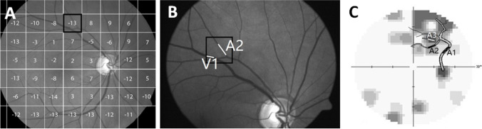
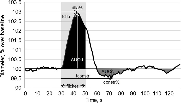
Methods: In patients with primary open angle glaucoma (POAG) (n = 30) and healthy controls (n = 22), NVC was studied using dynamic vessel analyzer to quantify retinal vessel diameter before, during, and after flicker light stimulation to evaluate the dilation response following neuronal activation. Vessel features and dilation were then related to branch level and visual field impairment.
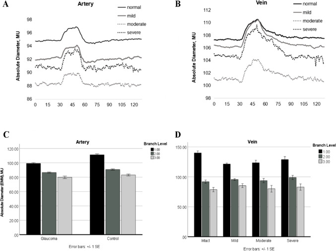
Results: Retinal arterial and venous vessels had significantly smaller diameters in patients with POAG in comparison to controls. However, both arterial and venous dilation reached normal values during neuronal activation despite their smaller diameters. This was largely independent of visual field depth and varied among patients.
Conclusions: Because dilation/constriction is normal, VD in POAG can be explained by chronic vasoconstriction which limits energy supply to retinal (and brain) neurons with subsequent hypo-metabolism ("silent" neurons) or neuronal cell death. We propose that the root cause of POAG is primarily of vascular and not neuronal origin. This understanding can help to better personalize POAG therapy of not only targeting eye pressure but also vasoconstriction to prevent low vision, slowing its progression and supporting recovery and restoration.
Trial registration: ClinicalTrials.gov, # NCT04037384 on July 3, 2019.
期刊介绍:
PMA Journal is a journal of predictive, preventive and personalized medicine (PPPM). The journal provides expert viewpoints and research on medical innovations and advanced healthcare using predictive diagnostics, targeted preventive measures and personalized patient treatments. The journal is indexed by PubMed, Embase and Scopus.


 分享
分享
 求助内容:
求助内容: 应助结果提醒方式:
 扫码关注我们
