Miesje M van der Stoel, Maria P Kotini, Rianne M Schoon, Markus Affolter, Heinz-Georg Belting, Stephan Huveneers
{"title":"Vinculin strengthens the endothelial barrier during vascular development.","authors":"Miesje M van der Stoel, Maria P Kotini, Rianne M Schoon, Markus Affolter, Heinz-Georg Belting, Stephan Huveneers","doi":"10.1530/VB-22-0012","DOIUrl":null,"url":null,"abstract":"<p><p>Remodelling of cell-cell junctions is crucial for proper tissue development and barrier function. The cadherin-based adherens junctions anchor via β-catenin and α-catenin to the actomyosin cytoskeleton, together forming a junctional mechanotransduction complex. Tension-induced conformational changes in the mechanosensitive α-catenin protein induce junctional vinculin recruitment. In endothelial cells, vinculin protects the remodelling of VE-cadherin junctions. In this study, we have addressed the role of vinculin in endothelial barrier function in the developing vasculature. In vitro experiments, using endothelial cells in which α-catenin was replaced by a vinculin-binding-deficient mutant, showed that junctional recruitment of vinculin promotes endothelial barrier function. To assess the role of vinculin within blood vessels in vivo, we next investigated barrier function in the vasculature of vcl knockout zebrafish. In the absence of vinculin, sprouting angiogenesis and vessel perfusion still occurred. Intriguingly, the absence of vinculin made the blood vessels more permeable for 10 kDa dextran molecules but not for larger tracers. Taken together, our findings demonstrate that vinculin strengthens the endothelial barrier and prevents vascular leakage in developing vessels.</p>","PeriodicalId":75294,"journal":{"name":"Vascular biology (Bristol, England)","volume":"5 1","pages":""},"PeriodicalIF":0.0000,"publicationDate":"2023-01-01","publicationTypes":"Journal Article","fieldsOfStudy":null,"isOpenAccess":false,"openAccessPdf":"https://ftp.ncbi.nlm.nih.gov/pub/pmc/oa_pdf/87/b0/VB-22-0012.PMC9986378.pdf","citationCount":"2","resultStr":null,"platform":"Semanticscholar","paperid":null,"PeriodicalName":"Vascular biology (Bristol, England)","FirstCategoryId":"1085","ListUrlMain":"https://doi.org/10.1530/VB-22-0012","RegionNum":0,"RegionCategory":null,"ArticlePicture":[],"TitleCN":null,"AbstractTextCN":null,"PMCID":null,"EPubDate":"","PubModel":"","JCR":"","JCRName":"","Score":null,"Total":0}
引用次数: 2
Abstract
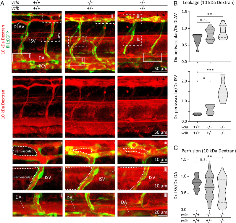
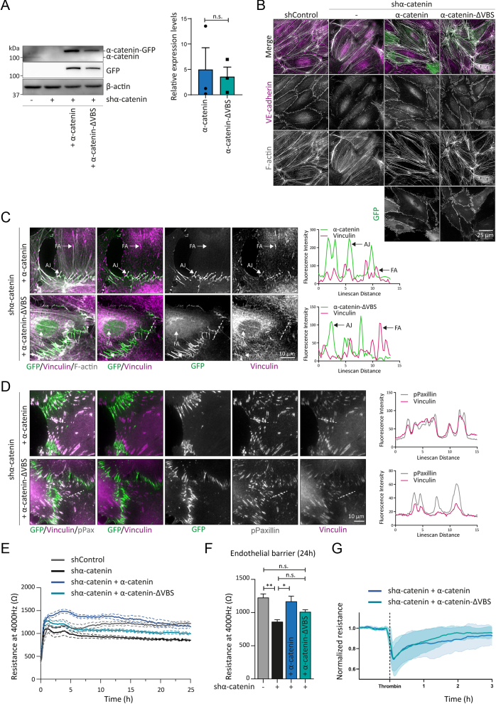
Remodelling of cell-cell junctions is crucial for proper tissue development and barrier function. The cadherin-based adherens junctions anchor via β-catenin and α-catenin to the actomyosin cytoskeleton, together forming a junctional mechanotransduction complex. Tension-induced conformational changes in the mechanosensitive α-catenin protein induce junctional vinculin recruitment. In endothelial cells, vinculin protects the remodelling of VE-cadherin junctions. In this study, we have addressed the role of vinculin in endothelial barrier function in the developing vasculature. In vitro experiments, using endothelial cells in which α-catenin was replaced by a vinculin-binding-deficient mutant, showed that junctional recruitment of vinculin promotes endothelial barrier function. To assess the role of vinculin within blood vessels in vivo, we next investigated barrier function in the vasculature of vcl knockout zebrafish. In the absence of vinculin, sprouting angiogenesis and vessel perfusion still occurred. Intriguingly, the absence of vinculin made the blood vessels more permeable for 10 kDa dextran molecules but not for larger tracers. Taken together, our findings demonstrate that vinculin strengthens the endothelial barrier and prevents vascular leakage in developing vessels.


 分享
分享

 求助内容:
求助内容: 应助结果提醒方式:
 扫码关注我们
