J.M. Plasencia Martínez , I. García Tuells , C. Bravo Pérez , A. Blanco Barrio
{"title":"Signo de la diana en COVID-19, significado radiológico y aportación diagnóstica de la tomosíntesis digital torácica","authors":"J.M. Plasencia Martínez , I. García Tuells , C. Bravo Pérez , A. Blanco Barrio","doi":"10.1016/j.rx.2023.07.006","DOIUrl":null,"url":null,"abstract":"<div><h3>Introduction</h3><p>Our objectives are to describe the imaging features, clinical characteristics, laboratory values and prognosis related to the target sign (TS) in COVID-19, as well as to determine whether digital tomosynthesis (DTS) of the chest has diagnostic advantages over radiography in this context.</p></div><div><h3>Material and methods</h3><p>Retrospective, descriptive, single-centre, case series study, accepted by our ethical committee. Radiological, clinical, analytical and follow-up characteristics of patients with COVID-19 and TS on radiography and DTS between November 2020 and January 2021 were analysed.</p></div><div><h3>Results</h3><p>Eleven TS were collected in seven patients, with a median age of 35 years, 57% male. All TS presented with a central nodule and a peripheral ring, and in at least 82%, the lung in between was of normal density. All TS were located in peripheral, basal regions and 91% in posterior regions. TS were multiple in 43%. The peripheral rings in contiguous TS were joined. Other pneumonia-related findings were identified in 86% of patients. Detection of the TS 82% higher in DTS than radiography. Only one patient underwent a CT pulmonary angiography, which was positive for acute pulmonary embolism. Seventy-one per cent presented with pleuritic pain. Laboratory values revealed no significant findings and the TS did not indicate a worse prognosis.</p></div><div><h3>Conclusions</h3><p>TS in COVID-19 predominates in peripheral and basal regions and can be multiple. Pulmonary embolism was detected in one case. It occurs in young people, frequently with pleuritic pain and does not lead to a worse prognosis. DTS detects TS in over 80% more cases than radiography.</p></div>","PeriodicalId":31509,"journal":{"name":"RADIOLOGIA","volume":"66 ","pages":"Pages S32-S39"},"PeriodicalIF":1.1000,"publicationDate":"2024-04-01","publicationTypes":"Journal Article","fieldsOfStudy":null,"isOpenAccess":false,"openAccessPdf":"","citationCount":"0","resultStr":null,"platform":"Semanticscholar","paperid":null,"PeriodicalName":"RADIOLOGIA","FirstCategoryId":"1085","ListUrlMain":"https://www.sciencedirect.com/science/article/pii/S0033833823001510","RegionNum":0,"RegionCategory":null,"ArticlePicture":[],"TitleCN":null,"AbstractTextCN":null,"PMCID":null,"EPubDate":"2023/8/28 0:00:00","PubModel":"Epub","JCR":"Q3","JCRName":"RADIOLOGY, NUCLEAR MEDICINE & MEDICAL IMAGING","Score":null,"Total":0}
引用次数: 0
Abstract
Introduction
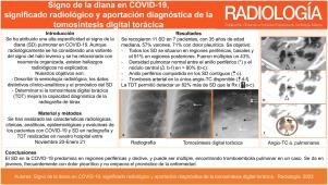
Our objectives are to describe the imaging features, clinical characteristics, laboratory values and prognosis related to the target sign (TS) in COVID-19, as well as to determine whether digital tomosynthesis (DTS) of the chest has diagnostic advantages over radiography in this context.
Material and methods
Retrospective, descriptive, single-centre, case series study, accepted by our ethical committee. Radiological, clinical, analytical and follow-up characteristics of patients with COVID-19 and TS on radiography and DTS between November 2020 and January 2021 were analysed.
Results
Eleven TS were collected in seven patients, with a median age of 35 years, 57% male. All TS presented with a central nodule and a peripheral ring, and in at least 82%, the lung in between was of normal density. All TS were located in peripheral, basal regions and 91% in posterior regions. TS were multiple in 43%. The peripheral rings in contiguous TS were joined. Other pneumonia-related findings were identified in 86% of patients. Detection of the TS 82% higher in DTS than radiography. Only one patient underwent a CT pulmonary angiography, which was positive for acute pulmonary embolism. Seventy-one per cent presented with pleuritic pain. Laboratory values revealed no significant findings and the TS did not indicate a worse prognosis.
Conclusions
TS in COVID-19 predominates in peripheral and basal regions and can be multiple. Pulmonary embolism was detected in one case. It occurs in young people, frequently with pleuritic pain and does not lead to a worse prognosis. DTS detects TS in over 80% more cases than radiography.
RADIOLOGIARADIOLOGY, NUCLEAR MEDICINE & MEDICAL IMAGING-
CiteScore
1.60
自引率
7.70%
发文量
105
审稿时长
52 days
期刊介绍:
La mejor revista para conocer de primera mano los originales más relevantes en la especialidad y las revisiones, casos y notas clínicas de mayor interés profesional. Además es la Publicación Oficial de la Sociedad Española de Radiología Médica.


 分享
分享
 求助内容:
求助内容: 应助结果提醒方式:
 扫码关注我们
