{"title":"SARS-CoV-2 Infection in Late Pregnancy and Childbirth from the Perspective of Perinatal Pathology.","authors":"Larisa Debelenko","doi":"10.3390/jdb11040042","DOIUrl":null,"url":null,"abstract":"<p><p>This review focuses on SARS-CoV-2 infection in placental and fetal tissues. Viremia is rare in infected pregnant women, and the virus is seldom amplified from placental tissues. Definite and probable placental infection requires the demonstration of viral RNA or proteins using in situ hybridization (ISH) and immunohistochemistry (IHC). Small subsets (1.0-7.9%, median 2.8%) of placentas of SARS-CoV-2-positive women showed definite infection accompanied by a characteristic histopathology named SARS-CoV-2 placentitis (SP). The conventionally accepted histopathological criteria for SP include the triad of intervillositis, perivillous fibrin deposition, and trophoblast necrosis. SP was shown to be independent of the clinical severity of the infection, but associated with stillbirth in cases where destructive lesions affecting more than 75% of the placental tissue resulted in placental insufficiency and severe fetal hypoxic-ischemic injury. An association between maternal thrombophilia and SP was shown in a subset of cases, suggesting a synergy of the infection and deficient coagulation cascade as one of the mechanisms of the pathologic accumulation of fibrin in affected placentas. The virus was amplified from fetal tissues in approximately 40% of SP cases, but definite fetal involvement demonstrated using ISH or IHC is exceptionally rare. The placental pathology in SARS-CoV-2-positive women also includes chronic lesions associated with placental malperfusion in the absence of definite or probable placental infection. The direct viral causation of the vascular malperfusion of the placenta in COVID-19 is debatable, and common predispositions (hypertension, diabetes, and obesity) may play a role.</p>","PeriodicalId":15563,"journal":{"name":"Journal of Developmental Biology","volume":"11 4","pages":""},"PeriodicalIF":2.5000,"publicationDate":"2023-11-16","publicationTypes":"Journal Article","fieldsOfStudy":null,"isOpenAccess":false,"openAccessPdf":"https://www.ncbi.nlm.nih.gov/pmc/articles/PMC10660738/pdf/","citationCount":"0","resultStr":null,"platform":"Semanticscholar","paperid":null,"PeriodicalName":"Journal of Developmental Biology","FirstCategoryId":"1085","ListUrlMain":"https://doi.org/10.3390/jdb11040042","RegionNum":0,"RegionCategory":null,"ArticlePicture":[],"TitleCN":null,"AbstractTextCN":null,"PMCID":null,"EPubDate":"","PubModel":"","JCR":"Q3","JCRName":"DEVELOPMENTAL BIOLOGY","Score":null,"Total":0}
引用次数: 0
Abstract
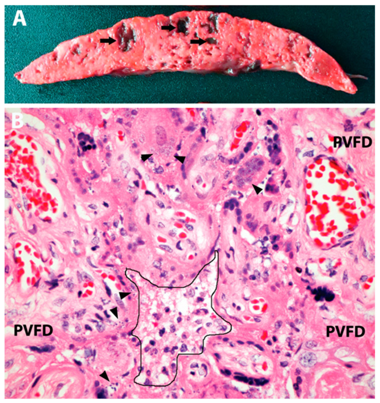
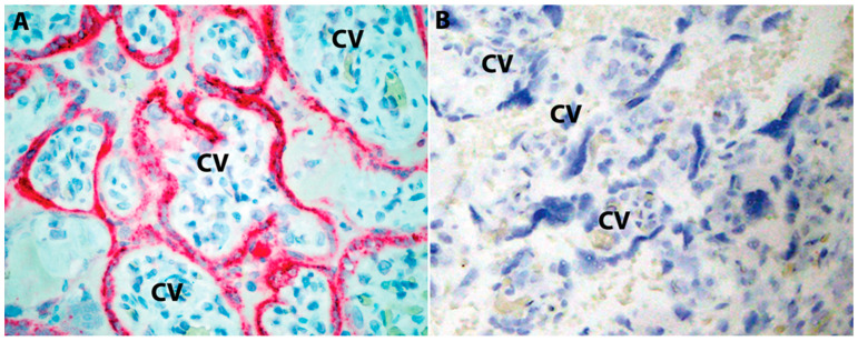
This review focuses on SARS-CoV-2 infection in placental and fetal tissues. Viremia is rare in infected pregnant women, and the virus is seldom amplified from placental tissues. Definite and probable placental infection requires the demonstration of viral RNA or proteins using in situ hybridization (ISH) and immunohistochemistry (IHC). Small subsets (1.0-7.9%, median 2.8%) of placentas of SARS-CoV-2-positive women showed definite infection accompanied by a characteristic histopathology named SARS-CoV-2 placentitis (SP). The conventionally accepted histopathological criteria for SP include the triad of intervillositis, perivillous fibrin deposition, and trophoblast necrosis. SP was shown to be independent of the clinical severity of the infection, but associated with stillbirth in cases where destructive lesions affecting more than 75% of the placental tissue resulted in placental insufficiency and severe fetal hypoxic-ischemic injury. An association between maternal thrombophilia and SP was shown in a subset of cases, suggesting a synergy of the infection and deficient coagulation cascade as one of the mechanisms of the pathologic accumulation of fibrin in affected placentas. The virus was amplified from fetal tissues in approximately 40% of SP cases, but definite fetal involvement demonstrated using ISH or IHC is exceptionally rare. The placental pathology in SARS-CoV-2-positive women also includes chronic lesions associated with placental malperfusion in the absence of definite or probable placental infection. The direct viral causation of the vascular malperfusion of the placenta in COVID-19 is debatable, and common predispositions (hypertension, diabetes, and obesity) may play a role.
期刊介绍:
The Journal of Developmental Biology (ISSN 2221-3759) is an international, peer-reviewed, quick-refereeing, open access journal, which publishes reviews, research papers and communications on the development of multicellular organisms at the molecule, cell, tissue, organ and whole organism levels. Our aim is to encourage researchers to effortlessly publish their new findings or concepts rapidly in an open access medium, overseen by their peers. There is no restriction on the length of the papers; the full experimental details must be provided so that the results can be reproduced. Electronic files regarding the full details of the experimental procedure, if unable to be published in a normal way, can be deposited as supplementary material. Journal of Developmental Biology focuses on: -Development mechanisms and genetics -Cell differentiation -Embryonal development -Tissue/organism growth -Metamorphosis and regeneration of the organisms. It involves many biological fields, such as Molecular biology, Genetics, Physiology, Cell biology, Anatomy, Embryology, Cancer research, Neurobiology, Immunology, Ecology, Evolutionary biology.


 分享
分享
 求助内容:
求助内容: 应助结果提醒方式:
 扫码关注我们
