Early Detection of hyperemia with Magnetic Resonance Fluid Attenuation Inversion Recovery Imaging after Superficial Temporal Artery to Middle Cerebral Artery Anastomosis.
{"title":"Early Detection of hyperemia with Magnetic Resonance Fluid Attenuation Inversion Recovery Imaging after Superficial Temporal Artery to Middle Cerebral Artery Anastomosis.","authors":"Jin Eun, Ik Seong Park","doi":"10.3340/jkns.2023.0183","DOIUrl":null,"url":null,"abstract":"<p><strong>Objective: </strong>Cerebral hyperperfusion syndrome (CHS) manifests as a collection of symptoms brought on by heightened focal cerebral blood flow (CBF), afflicting nearly 30% of patients who have undergone superficial temporal artery (STA)-middle cerebral artery (MCA) anastomosis. The aim of this study was to investigate whether the amalgamation of magnetic resonance imaging (MRI) fluid-attenuated inversion recovery (FLAIR) and apparent diffusion coefficient (ADC) imaging via MRI can discern cerebral hyperemia after STA-MCA anastomosis surgery.</p><p><strong>Methods: </strong>A retrospective study was performed of patients who underwent STA-MCA anastomosis due to Moyamoya disease or atherosclerotic steno-occlusive disease. A protocol aimed at preventing CHS was instituted, leveraging the use of MRI FLAIR. Patients underwent MRI diffusion with FLAIR imaging 24 hours after STA-MCA anastomosis. A high signal on FLAIR images signified the presence of hyperemia at the bypass site, triggering a protocol of hyperemia care. All patients underwent hemodynamic evaluations, including perfusion MRI, single-photon emission computed tomography (SPECT), and digital subtraction angiography, both before and after the surgery. If a high signal intensity is observed on MRI FLAIR within 24 hours of the surgery, a repeat MRI is performed to confirm the presence of hyperemia. Patients with confirmed hyperemia are managed according to a protocol aimed at preventing further progression.</p><p><strong>Results: </strong>Out of a total of 162 patients, 24 individuals (comprising 16 women and 8 men) exhibited hyperemia on their MRI FLAIR scans following the procedure. SPECT was conducted on 23 patients, and 11 of them yielded positive results. All 24 patients underwent perfusion MRI, but nine of them showed no significant findings. Among the patients, 10 displayed elevations in both CBF and cerebral blood volume (CBV), three only showed elevation in CBF, and two only showed elevation in CBV. Follow-up MRI FLAIR scans conducted 6 months later on these patients revealed complete normalization of the previously observed high signal intensity, with no evidence of ischemic injury.</p><p><strong>Conclusion: </strong>The study determined that the use of MRI FLAIR and ADC mapping is a competent means of early detection of hyperemia after STA-MCA anastomosis surgery. The protocol established can be adopted by other neurosurgical institutions to enhance patient outcomes and mitigate the hazard of permanent cerebral injury caused by cerebral hyperemia.</p>","PeriodicalId":16283,"journal":{"name":"Journal of Korean Neurosurgical Society","volume":" ","pages":"442-450"},"PeriodicalIF":1.7000,"publicationDate":"2024-07-01","publicationTypes":"Journal Article","fieldsOfStudy":null,"isOpenAccess":false,"openAccessPdf":"https://www.ncbi.nlm.nih.gov/pmc/articles/PMC11220419/pdf/","citationCount":"0","resultStr":null,"platform":"Semanticscholar","paperid":null,"PeriodicalName":"Journal of Korean Neurosurgical Society","FirstCategoryId":"3","ListUrlMain":"https://doi.org/10.3340/jkns.2023.0183","RegionNum":4,"RegionCategory":"医学","ArticlePicture":[],"TitleCN":null,"AbstractTextCN":null,"PMCID":null,"EPubDate":"2023/11/21 0:00:00","PubModel":"Epub","JCR":"Q4","JCRName":"CLINICAL NEUROLOGY","Score":null,"Total":0}
引用次数: 0
Abstract
Objective: Cerebral hyperperfusion syndrome (CHS) manifests as a collection of symptoms brought on by heightened focal cerebral blood flow (CBF), afflicting nearly 30% of patients who have undergone superficial temporal artery (STA)-middle cerebral artery (MCA) anastomosis. The aim of this study was to investigate whether the amalgamation of magnetic resonance imaging (MRI) fluid-attenuated inversion recovery (FLAIR) and apparent diffusion coefficient (ADC) imaging via MRI can discern cerebral hyperemia after STA-MCA anastomosis surgery.
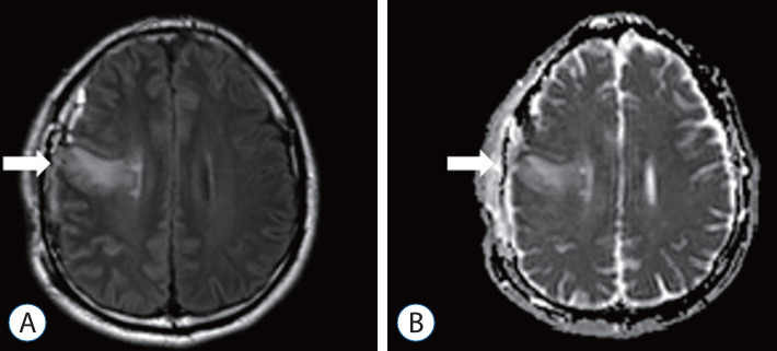
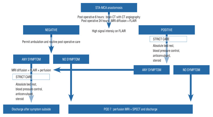
Methods: A retrospective study was performed of patients who underwent STA-MCA anastomosis due to Moyamoya disease or atherosclerotic steno-occlusive disease. A protocol aimed at preventing CHS was instituted, leveraging the use of MRI FLAIR. Patients underwent MRI diffusion with FLAIR imaging 24 hours after STA-MCA anastomosis. A high signal on FLAIR images signified the presence of hyperemia at the bypass site, triggering a protocol of hyperemia care. All patients underwent hemodynamic evaluations, including perfusion MRI, single-photon emission computed tomography (SPECT), and digital subtraction angiography, both before and after the surgery. If a high signal intensity is observed on MRI FLAIR within 24 hours of the surgery, a repeat MRI is performed to confirm the presence of hyperemia. Patients with confirmed hyperemia are managed according to a protocol aimed at preventing further progression.
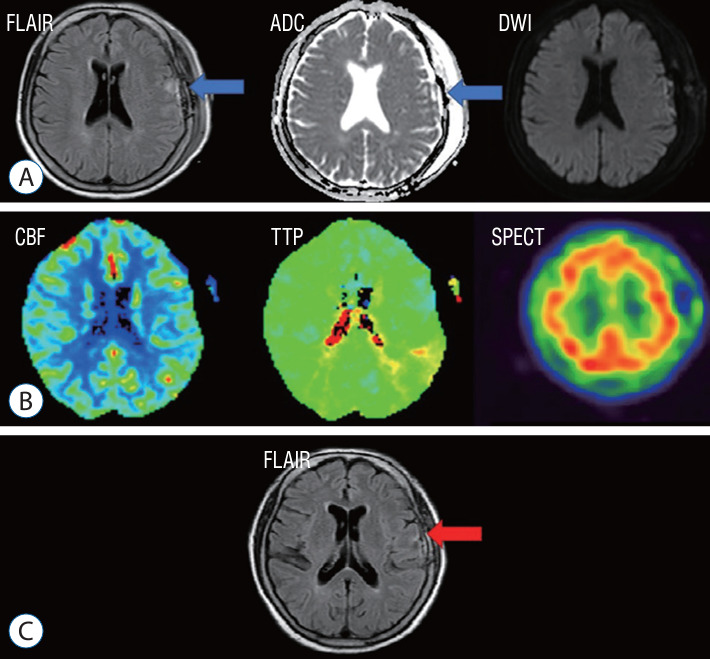
Results: Out of a total of 162 patients, 24 individuals (comprising 16 women and 8 men) exhibited hyperemia on their MRI FLAIR scans following the procedure. SPECT was conducted on 23 patients, and 11 of them yielded positive results. All 24 patients underwent perfusion MRI, but nine of them showed no significant findings. Among the patients, 10 displayed elevations in both CBF and cerebral blood volume (CBV), three only showed elevation in CBF, and two only showed elevation in CBV. Follow-up MRI FLAIR scans conducted 6 months later on these patients revealed complete normalization of the previously observed high signal intensity, with no evidence of ischemic injury.
Conclusion: The study determined that the use of MRI FLAIR and ADC mapping is a competent means of early detection of hyperemia after STA-MCA anastomosis surgery. The protocol established can be adopted by other neurosurgical institutions to enhance patient outcomes and mitigate the hazard of permanent cerebral injury caused by cerebral hyperemia.
期刊介绍:
The Journal of Korean Neurosurgical Society (J Korean Neurosurg Soc) is the official journal of the Korean Neurosurgical Society, and published bimonthly (1st day of January, March, May, July, September, and November). It launched in October 31, 1972 with Volume 1 and Number 1. J Korean Neurosurg Soc aims to allow neurosurgeons from around the world to enrich their knowledge of patient management, education, and clinical or experimental research, and hence their professionalism. This journal publishes Laboratory Investigations, Clinical Articles, Review Articles, Case Reports, Technical Notes, and Letters to the Editor. Our field of interest involves clinical neurosurgery (cerebrovascular disease, neuro-oncology, skull base neurosurgery, spine, pediatric neurosurgery, functional neurosurgery, epilepsy, neuro-trauma, and peripheral nerve disease) and laboratory work in neuroscience.


 分享
分享
 求助内容:
求助内容: 应助结果提醒方式:
 扫码关注我们
