Association between P-wave terminal force in lead V1 and extent of left atrial low-voltage substrate in older patients with paroxysmal atrial fibrillation.
{"title":"Association between P-wave terminal force in lead V<sub>1</sub> and extent of left atrial low-voltage substrate in older patients with paroxysmal atrial fibrillation.","authors":"Yue Qiu, Jinyu Sun, Yuxuan Wang, Caiyi Jin, Weizhu Ju, Gang Yang, Kai Gu, Hailei Liu, Zidun Wang, Xiaohong Jiang, Mingfang Li, Hongwu Chen, Minglong Chen","doi":"10.1007/s10840-023-01710-w","DOIUrl":null,"url":null,"abstract":"<p><strong>Background: </strong>The P-wave terminal force in lead V<sub>1</sub> (PTFV<sub>1</sub>) is a marker of cardiomyopathy and risk of atrial fibrillation (AF). Low-voltage area (LVA) in the left atrium (LA), which indicates underlying atrial fibrosis, could predict AF recurrence. This study aimed to investigate the correlation between PTFV<sub>1</sub> and LVA in older patients with paroxysmal AF.</p><p><strong>Methods: </strong>From May 1, 2020, to October 31, 2021, a total of 162 patients aged 65-80 years with paroxysmal AF who underwent index ablation procedures were enrolled. PTFV<sub>1</sub> was measured in sinus rhythm (SR) using 12-lead electrocardiograms prior to the ablation. Abnormal PTFV<sub>1</sub> was defined as a ≥ 4 mVms depression. Additional LVA ablation beyond circumferential pulmonary vein isolation (CPVI) was performed if LVAs were found.</p><p><strong>Results: </strong>Among the 162 patients, 88 had a normal PTFV<sub>1</sub> and 74 had an abnormal PTFV<sub>1</sub> prior to ablation. There was a significant difference in LVA in patients with and without an abnormal PTFV<sub>1</sub> (LVA, 11.0 vs. 5.1 cm<sup>2</sup>, P < 0.001; LVA burden, 8.9% vs. 4.5%, P < 0.001). PTFV<sub>1</sub> and PTAV<sub>1</sub> were highest in the upper tertile with extensive LVAs (P < 0.001). Multivariate analysis revealed that abnormal PTFV<sub>1</sub> was an independent predictor of LVAs (β = 4.961; 95% CI, 2.135-7.788; P < 0.001). After a median follow-up of 23 months, the AF-free survival rate was similar between the normal PTFV<sub>1</sub> group and the abnormal PTFV<sub>1</sub> group (13/88 vs. 12/74, hazard ratio [HR], 0.933 [95% CI, 0.425-2.047]; P = 0.861).</p><p><strong>Conclusions: </strong>Abnormal PTFV<sub>1</sub> at baseline was independently associated with the extent of LVA in older patients with paroxysmal AF.</p>","PeriodicalId":16202,"journal":{"name":"Journal of Interventional Cardiac Electrophysiology","volume":" ","pages":"1153-1160"},"PeriodicalIF":2.6000,"publicationDate":"2024-08-01","publicationTypes":"Journal Article","fieldsOfStudy":null,"isOpenAccess":false,"openAccessPdf":"","citationCount":"0","resultStr":null,"platform":"Semanticscholar","paperid":null,"PeriodicalName":"Journal of Interventional Cardiac Electrophysiology","FirstCategoryId":"3","ListUrlMain":"https://doi.org/10.1007/s10840-023-01710-w","RegionNum":4,"RegionCategory":"医学","ArticlePicture":[],"TitleCN":null,"AbstractTextCN":null,"PMCID":null,"EPubDate":"2023/11/30 0:00:00","PubModel":"Epub","JCR":"Q3","JCRName":"CARDIAC & CARDIOVASCULAR SYSTEMS","Score":null,"Total":0}
引用次数: 0
Abstract
Background: The P-wave terminal force in lead V1 (PTFV1) is a marker of cardiomyopathy and risk of atrial fibrillation (AF). Low-voltage area (LVA) in the left atrium (LA), which indicates underlying atrial fibrosis, could predict AF recurrence. This study aimed to investigate the correlation between PTFV1 and LVA in older patients with paroxysmal AF.
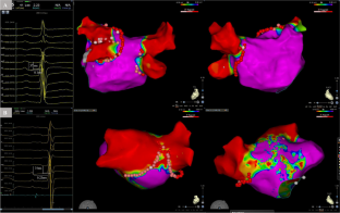
Methods: From May 1, 2020, to October 31, 2021, a total of 162 patients aged 65-80 years with paroxysmal AF who underwent index ablation procedures were enrolled. PTFV1 was measured in sinus rhythm (SR) using 12-lead electrocardiograms prior to the ablation. Abnormal PTFV1 was defined as a ≥ 4 mVms depression. Additional LVA ablation beyond circumferential pulmonary vein isolation (CPVI) was performed if LVAs were found.
Results: Among the 162 patients, 88 had a normal PTFV1 and 74 had an abnormal PTFV1 prior to ablation. There was a significant difference in LVA in patients with and without an abnormal PTFV1 (LVA, 11.0 vs. 5.1 cm2, P < 0.001; LVA burden, 8.9% vs. 4.5%, P < 0.001). PTFV1 and PTAV1 were highest in the upper tertile with extensive LVAs (P < 0.001). Multivariate analysis revealed that abnormal PTFV1 was an independent predictor of LVAs (β = 4.961; 95% CI, 2.135-7.788; P < 0.001). After a median follow-up of 23 months, the AF-free survival rate was similar between the normal PTFV1 group and the abnormal PTFV1 group (13/88 vs. 12/74, hazard ratio [HR], 0.933 [95% CI, 0.425-2.047]; P = 0.861).
Conclusions: Abnormal PTFV1 at baseline was independently associated with the extent of LVA in older patients with paroxysmal AF.
期刊介绍:
The Journal of Interventional Cardiac Electrophysiology is an international publication devoted to fostering research in and development of interventional techniques and therapies for the management of cardiac arrhythmias. It is designed primarily to present original research studies and scholarly scientific reviews of basic and applied science and clinical research in this field. The Journal will adopt a multidisciplinary approach to link physical, experimental, and clinical sciences as applied to the development of and practice in interventional electrophysiology. The Journal will examine techniques ranging from molecular, chemical and pharmacologic therapies to device and ablation technology. Accordingly, original research in clinical, epidemiologic and basic science arenas will be considered for publication. Applied engineering or physical science studies pertaining to interventional electrophysiology will be encouraged. The Journal is committed to providing comprehensive and detailed treatment of major interventional therapies and innovative techniques in a structured and clinically relevant manner. It is directed at clinical practitioners and investigators in the rapidly growing field of interventional electrophysiology. The editorial staff and board reflect this bias and include noted international experts in this area with a wealth of expertise in basic and clinical investigation. Peer review of all submissions, conflict of interest guidelines and periodic editorial board review of all Journal policies have been established.


 分享
分享
 求助内容:
求助内容: 应助结果提醒方式:
 扫码关注我们
