Nei Fukasawa, Miku Maeda, Yoshifumi Sugiyama, Takahiro Fukuda, Masayuki Shimoda
{"title":"Distribution of proteinase K-resistant anti-α-synuclein immunoreactive axons in the cardiac plexus is unbiased to the left ventricular anterior wall.","authors":"Nei Fukasawa, Miku Maeda, Yoshifumi Sugiyama, Takahiro Fukuda, Masayuki Shimoda","doi":"10.1111/pin.13389","DOIUrl":null,"url":null,"abstract":"<p><p>Lewy body disease (LBD) is characterized by the appearance of Lewy neurites and Lewy bodies, which are predominantly composed of α-synuclein. Notably, the cardiac plexus (CP) is one of the main targets of LBD research. Although previous studies have reported obvious differences in the frequency of Lewy body pathology (LBP) in the CP, none of them have confirmed whether LBP preferably appears in any part of the CP. Thus, we aimed to clarify the emergence and/or propagation of LBP in the CP. In this study, 263 consecutive autopsy cases of patients aged ≥50 years were included, with one region per case selected from three myocardial perfusion areas (MPAs) and subjected to proteinase K and then immunohistochemically stained with anti-α-synuclein antibodies to assess LBP. We stained all three MPAs in 17 cases with low-density LBP and observed the actual distribution of LBP. LBP were identified in the CP in 20.2% (53/263) of patients. Moreover, we found that LBP may appear in only one region of MPAs, mainly in the young-old group (35.3% (6/17) of patients). These findings suggest that it is possible to underestimate LBP in the CP, especially in the young-old group, by restricting the search to only one of the three MPAs.</p>","PeriodicalId":19806,"journal":{"name":"Pathology International","volume":" ","pages":"1-12"},"PeriodicalIF":3.4000,"publicationDate":"2024-01-01","publicationTypes":"Journal Article","fieldsOfStudy":null,"isOpenAccess":false,"openAccessPdf":"https://www.ncbi.nlm.nih.gov/pmc/articles/PMC11551827/pdf/","citationCount":"0","resultStr":null,"platform":"Semanticscholar","paperid":null,"PeriodicalName":"Pathology International","FirstCategoryId":"3","ListUrlMain":"https://doi.org/10.1111/pin.13389","RegionNum":4,"RegionCategory":"医学","ArticlePicture":[],"TitleCN":null,"AbstractTextCN":null,"PMCID":null,"EPubDate":"2023/12/1 0:00:00","PubModel":"Epub","JCR":"Q2","JCRName":"PATHOLOGY","Score":null,"Total":0}
引用次数: 0
Abstract
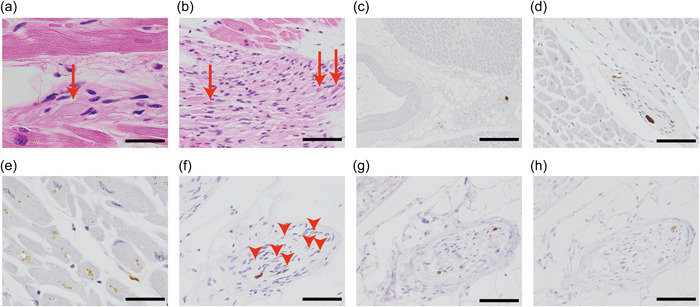
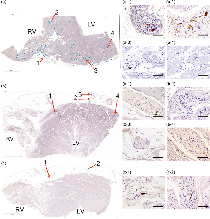
Lewy body disease (LBD) is characterized by the appearance of Lewy neurites and Lewy bodies, which are predominantly composed of α-synuclein. Notably, the cardiac plexus (CP) is one of the main targets of LBD research. Although previous studies have reported obvious differences in the frequency of Lewy body pathology (LBP) in the CP, none of them have confirmed whether LBP preferably appears in any part of the CP. Thus, we aimed to clarify the emergence and/or propagation of LBP in the CP. In this study, 263 consecutive autopsy cases of patients aged ≥50 years were included, with one region per case selected from three myocardial perfusion areas (MPAs) and subjected to proteinase K and then immunohistochemically stained with anti-α-synuclein antibodies to assess LBP. We stained all three MPAs in 17 cases with low-density LBP and observed the actual distribution of LBP. LBP were identified in the CP in 20.2% (53/263) of patients. Moreover, we found that LBP may appear in only one region of MPAs, mainly in the young-old group (35.3% (6/17) of patients). These findings suggest that it is possible to underestimate LBP in the CP, especially in the young-old group, by restricting the search to only one of the three MPAs.
期刊介绍:
Pathology International is the official English journal of the Japanese Society of Pathology, publishing articles of excellence in human and experimental pathology. The Journal focuses on the morphological study of the disease process and/or mechanisms. For human pathology, morphological investigation receives priority but manuscripts describing the result of any ancillary methods (cellular, chemical, immunological and molecular biological) that complement the morphology are accepted. Manuscript on experimental pathology that approach pathologenesis or mechanisms of disease processes are expected to report on the data obtained from models using cellular, biochemical, molecular biological, animal, immunological or other methods in conjunction with morphology. Manuscripts that report data on laboratory medicine (clinical pathology) without significant morphological contribution are not accepted.


 分享
分享
 求助内容:
求助内容: 应助结果提醒方式:
 扫码关注我们
