Jakub Batko, Rafał Jakiel, Agata Krawczyk–Ożóg, Radosław Litwinowicz, Jakub Hołda, Stanisław Bartuś, Krzysztof Bartuś, Mateusz K. Hołda, Małgorzata Konieczyńska
{"title":"Definition and anatomical description of the left atrial appendage neck","authors":"Jakub Batko, Rafał Jakiel, Agata Krawczyk–Ożóg, Radosław Litwinowicz, Jakub Hołda, Stanisław Bartuś, Krzysztof Bartuś, Mateusz K. Hołda, Małgorzata Konieczyńska","doi":"10.1002/ca.24125","DOIUrl":null,"url":null,"abstract":"<p>The left atrial appendage (LAA) is well known as a source of cardiac thrombus formation. Despite its clinical importance, the LAA neck is still anatomically poorly defined. Therefore, this study aimed to define the LAA neck and determine its morphometric characteristics. We performed three-dimensional reconstructions of the heart chambers based on contrast-enhanced electrocardiography–gated computed tomography scans of 200 patients (47% females, 66.5 ± 13.6 years old). The LAA neck was defined as a truncated cone-shaped canal bounded proximally by the LAA orifice and distally by the lobe origin and was present in 98.0% of cases. The central axis of the LAA neck was 14.7 ± 2.3 mm. The mean area of the LAA neck walls was 856.6 ± 316.7 mm<sup>2</sup>. The LAA neck can be divided into aortic, arterial (the smallest), venous (the largest), and free surfaces. All areas have a trapezoidal shape with a broader proximal base. There were no statistically significant differences in the morphometric characteristics of the LAA neck between LAA types. Statistically significant differences between the sexes in the main morphometric parameters of the LAA neck were found in the central axis length and the LAA neck wall area. The LAA neck can be evaluated from computed tomography scans and their three-dimensional reconstructions. The current study provides a complex morphometric analysis of the LAA neck. The precise definition and morphometric details of the LAA neck presented in this study may influence the effectiveness and safety of LAA exclusion procedures.</p>","PeriodicalId":50687,"journal":{"name":"Clinical Anatomy","volume":"37 2","pages":"201-209"},"PeriodicalIF":2.3000,"publicationDate":"2023-11-29","publicationTypes":"Journal Article","fieldsOfStudy":null,"isOpenAccess":false,"openAccessPdf":"https://onlinelibrary.wiley.com/doi/epdf/10.1002/ca.24125","citationCount":"0","resultStr":null,"platform":"Semanticscholar","paperid":null,"PeriodicalName":"Clinical Anatomy","FirstCategoryId":"3","ListUrlMain":"https://onlinelibrary.wiley.com/doi/10.1002/ca.24125","RegionNum":4,"RegionCategory":"医学","ArticlePicture":[],"TitleCN":null,"AbstractTextCN":null,"PMCID":null,"EPubDate":"","PubModel":"","JCR":"Q1","JCRName":"ANATOMY & MORPHOLOGY","Score":null,"Total":0}
引用次数: 0
Abstract
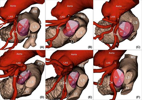
The left atrial appendage (LAA) is well known as a source of cardiac thrombus formation. Despite its clinical importance, the LAA neck is still anatomically poorly defined. Therefore, this study aimed to define the LAA neck and determine its morphometric characteristics. We performed three-dimensional reconstructions of the heart chambers based on contrast-enhanced electrocardiography–gated computed tomography scans of 200 patients (47% females, 66.5 ± 13.6 years old). The LAA neck was defined as a truncated cone-shaped canal bounded proximally by the LAA orifice and distally by the lobe origin and was present in 98.0% of cases. The central axis of the LAA neck was 14.7 ± 2.3 mm. The mean area of the LAA neck walls was 856.6 ± 316.7 mm2. The LAA neck can be divided into aortic, arterial (the smallest), venous (the largest), and free surfaces. All areas have a trapezoidal shape with a broader proximal base. There were no statistically significant differences in the morphometric characteristics of the LAA neck between LAA types. Statistically significant differences between the sexes in the main morphometric parameters of the LAA neck were found in the central axis length and the LAA neck wall area. The LAA neck can be evaluated from computed tomography scans and their three-dimensional reconstructions. The current study provides a complex morphometric analysis of the LAA neck. The precise definition and morphometric details of the LAA neck presented in this study may influence the effectiveness and safety of LAA exclusion procedures.
期刊介绍:
Clinical Anatomy is the Official Journal of the American Association of Clinical Anatomists and the British Association of Clinical Anatomists. The goal of Clinical Anatomy is to provide a medium for the exchange of current information between anatomists and clinicians. This journal embraces anatomy in all its aspects as applied to medical practice. Furthermore, the journal assists physicians and other health care providers in keeping abreast of new methodologies for patient management and informs educators of new developments in clinical anatomy and teaching techniques. Clinical Anatomy publishes original and review articles of scientific, clinical, and educational interest. Papers covering the application of anatomic principles to the solution of clinical problems and/or the application of clinical observations to expand anatomic knowledge are welcomed.


 分享
分享
 求助内容:
求助内容: 应助结果提醒方式:
 扫码关注我们
