Nadezhda V Kalacheva, Talia T Ginanova, Yaroslav O Kamenev, Sergey I Maslennikov, Igor Yu Dolmatov
{"title":"Morphology and ultrastructure of digestive system in pre-zoea and zoea I larvae of red king crab, Paralithodes camtschaticus (Tilesius, 1815).","authors":"Nadezhda V Kalacheva, Talia T Ginanova, Yaroslav O Kamenev, Sergey I Maslennikov, Igor Yu Dolmatov","doi":"10.1007/s00441-023-03843-w","DOIUrl":null,"url":null,"abstract":"<p><p>The digestive system structure in pre-zoea and zoea I larvae of the red king crab Paralithodes camtschaticus has been examined. During this development period, the digestive system consists of an esophagus, a stomach, a midgut (where the hepatopancreas ducts open), and a hindgut. The esophagus begins from the oral slit on the animal's ventral side and extends vertically up to the junction with the cardiac stomach. The latter is followed by the pyloric stomach. At the stages under study, crabs have a cardiac-pyloric valve and a pyloric filter in the stomach already developed. The midgut begins with an expansion in the cephalothorax, enters the pleon, grows narrower there, and extends to somite 3 of pleon. The hepatopancreas is represented by a symmetrical paired gland which occupies almost the entire cephalothorax space and opens with its ducts at the junction of the pyloric stomach with the midgut. The hepatopancreas is divided into the anterior and posterior lobes. At the pre-zoea stage, the anterior lobes are large and filled with yolk. At the zoea I stage, the anterior lobes are smaller relative to the entire hepatopancreas, and the posterior lobes increase and form tubular outgrowths. It has been shown that during the transition from pre-zoea to zoea I, the number of mitochondria in enterocytes increases and a peritrophic membrane forms in the midgut. These changes are probably associated with the transition to independent living and feeding.</p>","PeriodicalId":9712,"journal":{"name":"Cell and Tissue Research","volume":" ","pages":"1-20"},"PeriodicalIF":2.9000,"publicationDate":"2024-01-01","publicationTypes":"Journal Article","fieldsOfStudy":null,"isOpenAccess":false,"openAccessPdf":"","citationCount":"0","resultStr":null,"platform":"Semanticscholar","paperid":null,"PeriodicalName":"Cell and Tissue Research","FirstCategoryId":"99","ListUrlMain":"https://doi.org/10.1007/s00441-023-03843-w","RegionNum":3,"RegionCategory":"生物学","ArticlePicture":[],"TitleCN":null,"AbstractTextCN":null,"PMCID":null,"EPubDate":"2023/12/2 0:00:00","PubModel":"Epub","JCR":"Q3","JCRName":"CELL BIOLOGY","Score":null,"Total":0}
引用次数: 0
Abstract
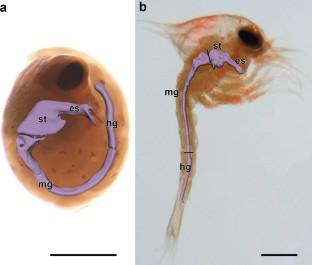
The digestive system structure in pre-zoea and zoea I larvae of the red king crab Paralithodes camtschaticus has been examined. During this development period, the digestive system consists of an esophagus, a stomach, a midgut (where the hepatopancreas ducts open), and a hindgut. The esophagus begins from the oral slit on the animal's ventral side and extends vertically up to the junction with the cardiac stomach. The latter is followed by the pyloric stomach. At the stages under study, crabs have a cardiac-pyloric valve and a pyloric filter in the stomach already developed. The midgut begins with an expansion in the cephalothorax, enters the pleon, grows narrower there, and extends to somite 3 of pleon. The hepatopancreas is represented by a symmetrical paired gland which occupies almost the entire cephalothorax space and opens with its ducts at the junction of the pyloric stomach with the midgut. The hepatopancreas is divided into the anterior and posterior lobes. At the pre-zoea stage, the anterior lobes are large and filled with yolk. At the zoea I stage, the anterior lobes are smaller relative to the entire hepatopancreas, and the posterior lobes increase and form tubular outgrowths. It has been shown that during the transition from pre-zoea to zoea I, the number of mitochondria in enterocytes increases and a peritrophic membrane forms in the midgut. These changes are probably associated with the transition to independent living and feeding.
期刊介绍:
The journal publishes regular articles and reviews in the areas of molecular, cell, and supracellular biology. In particular, the journal intends to provide a forum for publishing data that analyze the supracellular, integrative actions of gene products and their impact on the formation of tissue structure and function. Submission of papers with an emphasis on structure-function relationships as revealed by recombinant molecular technologies is especially encouraged. Areas of research with a long-standing tradition of publishing in Cell & Tissue Research include:
- neurobiology
- neuroendocrinology
- endocrinology
- reproductive biology
- skeletal and immune systems
- development
- stem cells
- muscle biology.


 分享
分享
 求助内容:
求助内容: 应助结果提醒方式:
 扫码关注我们
