{"title":"Deep learning approach for discrimination of liver lesions using nine time-phase images of contrast-enhanced ultrasound.","authors":"Naohisa Kamiyama, Katsutoshi Sugimoto, Ryuichi Nakahara, Tatsuya Kakegawa, Takao Itoi","doi":"10.1007/s10396-023-01390-z","DOIUrl":null,"url":null,"abstract":"<p><strong>Purpose: </strong>Contrast-enhanced ultrasound (CEUS) shows different enhancement patterns depending on the time after administration of the contrast agent. The aim of this study was to evaluate the diagnostic performance of liver nodule characterization using our proposed deep learning model with input of nine CEUS images.</p><p><strong>Methods: </strong>A total of 181 liver lesions (48 benign, 78 hepatocellular carcinoma (HCC), and 55 non-HCC malignant) were included in this prospective study. CEUS were performed using the contrast agent Sonazoid, and in addition to B-mode images before injection, image clips were stored every minute up to 10 min. A deep learning model was developed by arranging three ResNet50 transfer learning models in parallel. This proposed model allowed inputting up to nine datasets of different phases of CEUS and performing image augmentation of nine images synchronously. Using the results, the correct prediction rate, sensitivity, and specificity between \"benign\" and \"malignant\" cases were analyzed for each combination of the time phase. These accuracy values were also compared with the washout score judged by a human.</p><p><strong>Results: </strong>The proposed model showed performance superior to the referential standard model when the dataset from B-mode to the 10-min images were used (sensitivity: 93.2%, specificity: 65.3%, average correct answer rate: 60.1%). It also maintained 90.2% sensitivity and 61.2% specificity even when the dataset was limited to 2 min after injection, and this accuracy was equivalent to or better than human scoring by experts.</p><p><strong>Conclusion: </strong>Our proposed model has the potential to identify tumor types earlier than the Kupffer phase, but at the same time, machine learning confirmed that Kupffer-phase Sonazoid images contain essential information for the classification of liver nodules.</p>","PeriodicalId":50130,"journal":{"name":"Journal of Medical Ultrasonics","volume":" ","pages":"83-93"},"PeriodicalIF":2.1000,"publicationDate":"2024-01-01","publicationTypes":"Journal Article","fieldsOfStudy":null,"isOpenAccess":false,"openAccessPdf":"https://www.ncbi.nlm.nih.gov/pmc/articles/PMC12000262/pdf/","citationCount":"0","resultStr":null,"platform":"Semanticscholar","paperid":null,"PeriodicalName":"Journal of Medical Ultrasonics","FirstCategoryId":"3","ListUrlMain":"https://doi.org/10.1007/s10396-023-01390-z","RegionNum":4,"RegionCategory":"医学","ArticlePicture":[],"TitleCN":null,"AbstractTextCN":null,"PMCID":null,"EPubDate":"2023/12/5 0:00:00","PubModel":"Epub","JCR":"Q3","JCRName":"RADIOLOGY, NUCLEAR MEDICINE & MEDICAL IMAGING","Score":null,"Total":0}
引用次数: 0
Abstract
Purpose: Contrast-enhanced ultrasound (CEUS) shows different enhancement patterns depending on the time after administration of the contrast agent. The aim of this study was to evaluate the diagnostic performance of liver nodule characterization using our proposed deep learning model with input of nine CEUS images.
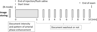
Methods: A total of 181 liver lesions (48 benign, 78 hepatocellular carcinoma (HCC), and 55 non-HCC malignant) were included in this prospective study. CEUS were performed using the contrast agent Sonazoid, and in addition to B-mode images before injection, image clips were stored every minute up to 10 min. A deep learning model was developed by arranging three ResNet50 transfer learning models in parallel. This proposed model allowed inputting up to nine datasets of different phases of CEUS and performing image augmentation of nine images synchronously. Using the results, the correct prediction rate, sensitivity, and specificity between "benign" and "malignant" cases were analyzed for each combination of the time phase. These accuracy values were also compared with the washout score judged by a human.
Results: The proposed model showed performance superior to the referential standard model when the dataset from B-mode to the 10-min images were used (sensitivity: 93.2%, specificity: 65.3%, average correct answer rate: 60.1%). It also maintained 90.2% sensitivity and 61.2% specificity even when the dataset was limited to 2 min after injection, and this accuracy was equivalent to or better than human scoring by experts.
Conclusion: Our proposed model has the potential to identify tumor types earlier than the Kupffer phase, but at the same time, machine learning confirmed that Kupffer-phase Sonazoid images contain essential information for the classification of liver nodules.
期刊介绍:
The Journal of Medical Ultrasonics is the official journal of the Japan Society of Ultrasonics in Medicine. The main purpose of the journal is to provide forum for the publication of papers documenting recent advances and new developments in the entire field of ultrasound in medicine and biology, encompassing both the medical and the engineering aspects of the science.The journal welcomes original articles, review articles, images, and letters to the editor.The journal also provides state-of-the-art information such as announcements from the boards and the committees of the society.


 分享
分享
 求助内容:
求助内容: 应助结果提醒方式:
 扫码关注我们
