{"title":"Tissue processing of endoscopic ultrasound-guided fine-needle aspiration specimens from solid pancreatic lesions.","authors":"Kenji Notohara, Kaori Nakamura","doi":"10.1007/s10396-023-01387-8","DOIUrl":null,"url":null,"abstract":"<p><p>Now that tissue cores can be obtained using fine-needle biopsy (FNB) needles, the ways tissues are handled for endoscopic ultrasound-guided fine-needle aspiration (EUS-FNA) are changing. Direct smear, touch smear of core tissues, and centrifugation have been used for cytological examinations, and liquid-based cytology (LBC), which allows immunostaining and genetic tests that use residual samples, is emerging as an alternative. We emphasize that liquid cytology (Cytospin™ cytology and LBC) is still important, because it enables the diagnosis of pancreatic ductal adenocarcinoma (PDAC) when cancerous cells are scarce in specimens. Cell blocks are being replaced by core tissues obtained via FNB needles. Recent reports indicate that rapid on-site evaluation (ROSE) is not necessary when FNB needles are used, and macroscopic on-site evaluation is used to evaluate specimen adequacy. Macroscopic findings of specimens are helpful in the diagnostic workup and for clarifying specimen-handling methods. In addition to the red strings and white cores observed in PDAC, mixed red and white strings, gray tissues, and gelatinous tissues are observed. Gray (necrotic) tissues and gelatinous (mucus) tissues are more suitable than histology for cell block or cytological processing. Tumor cells in neuroendocrine tumors (NETs) are numerous in red strings but cannot be observed macroscopically. ROSE might thus be necessary for lesions that may be NETs. Core tissues can be used for genetic tests, such as those used for KRAS mutations and comprehensive genomic profiling. Cytological materials, including slides and LBC specimens, can also be genetic test materials.</p>","PeriodicalId":50130,"journal":{"name":"Journal of Medical Ultrasonics","volume":" ","pages":"261-274"},"PeriodicalIF":2.1000,"publicationDate":"2024-04-01","publicationTypes":"Journal Article","fieldsOfStudy":null,"isOpenAccess":false,"openAccessPdf":"","citationCount":"0","resultStr":null,"platform":"Semanticscholar","paperid":null,"PeriodicalName":"Journal of Medical Ultrasonics","FirstCategoryId":"3","ListUrlMain":"https://doi.org/10.1007/s10396-023-01387-8","RegionNum":4,"RegionCategory":"医学","ArticlePicture":[],"TitleCN":null,"AbstractTextCN":null,"PMCID":null,"EPubDate":"2023/12/5 0:00:00","PubModel":"Epub","JCR":"Q3","JCRName":"RADIOLOGY, NUCLEAR MEDICINE & MEDICAL IMAGING","Score":null,"Total":0}
引用次数: 0
Abstract
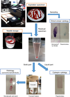
Now that tissue cores can be obtained using fine-needle biopsy (FNB) needles, the ways tissues are handled for endoscopic ultrasound-guided fine-needle aspiration (EUS-FNA) are changing. Direct smear, touch smear of core tissues, and centrifugation have been used for cytological examinations, and liquid-based cytology (LBC), which allows immunostaining and genetic tests that use residual samples, is emerging as an alternative. We emphasize that liquid cytology (Cytospin™ cytology and LBC) is still important, because it enables the diagnosis of pancreatic ductal adenocarcinoma (PDAC) when cancerous cells are scarce in specimens. Cell blocks are being replaced by core tissues obtained via FNB needles. Recent reports indicate that rapid on-site evaluation (ROSE) is not necessary when FNB needles are used, and macroscopic on-site evaluation is used to evaluate specimen adequacy. Macroscopic findings of specimens are helpful in the diagnostic workup and for clarifying specimen-handling methods. In addition to the red strings and white cores observed in PDAC, mixed red and white strings, gray tissues, and gelatinous tissues are observed. Gray (necrotic) tissues and gelatinous (mucus) tissues are more suitable than histology for cell block or cytological processing. Tumor cells in neuroendocrine tumors (NETs) are numerous in red strings but cannot be observed macroscopically. ROSE might thus be necessary for lesions that may be NETs. Core tissues can be used for genetic tests, such as those used for KRAS mutations and comprehensive genomic profiling. Cytological materials, including slides and LBC specimens, can also be genetic test materials.
期刊介绍:
The Journal of Medical Ultrasonics is the official journal of the Japan Society of Ultrasonics in Medicine. The main purpose of the journal is to provide forum for the publication of papers documenting recent advances and new developments in the entire field of ultrasound in medicine and biology, encompassing both the medical and the engineering aspects of the science.The journal welcomes original articles, review articles, images, and letters to the editor.The journal also provides state-of-the-art information such as announcements from the boards and the committees of the society.


 分享
分享
 求助内容:
求助内容: 应助结果提醒方式:
 扫码关注我们
