{"title":"DISTAL FEMUR HEMIEPIPHYSIODESIS IN KNEE RECURVATUM: A NEW SURGICAL TECHNIQUE.","authors":"Nei Botter Montenegro, Talissa Oliveira Generoso, Bárbara Lívia Corrêa Serafim, Amâncio Ramalho","doi":"10.1590/1413-785220233105e268307","DOIUrl":null,"url":null,"abstract":"<p><strong>Introduction: </strong>The genu recurvatum is characterized by a hyperextension deformity of the knee in the sagittal plane. Among its causes are conditions such as arthrogryposis, cerebral palsy, poliomyelitis, sequelae of tibial tuberosity fracture and some syndromes with generalized joint hypermobility. Treatment of this deformity can be challenging and, to date, aggressive methods such as femur or tibial osteotomies are the most used for its correction.</p><p><strong>Objective: </strong>This study aimed to describe a new surgical technique for correcting genu recurvatum.</p><p><strong>Methods: </strong>This is a prospective clinical study of children who underwent posterior hemiepiphysiodesis of the distal femur with transphyseal screws.</p><p><strong>Results: </strong>The approach proved to be safe and useful for genu recurvatum deformities, with femoral or articular apex.</p><p><strong>Conclusion: </strong>This approach shows great potential for correcting genu recurvatum in the developing skeleton, being an excellent alternative to the more aggressive methods currently used to treat this deformity. <b><i>Level of evidence IV, Case Series.</i></b></p>","PeriodicalId":55563,"journal":{"name":"Acta Ortopedica Brasileira","volume":"31 6","pages":"e268307"},"PeriodicalIF":0.6000,"publicationDate":"2023-12-18","publicationTypes":"Journal Article","fieldsOfStudy":null,"isOpenAccess":false,"openAccessPdf":"https://www.ncbi.nlm.nih.gov/pmc/articles/PMC10726712/pdf/","citationCount":"0","resultStr":null,"platform":"Semanticscholar","paperid":null,"PeriodicalName":"Acta Ortopedica Brasileira","FirstCategoryId":"3","ListUrlMain":"https://doi.org/10.1590/1413-785220233105e268307","RegionNum":4,"RegionCategory":"医学","ArticlePicture":[],"TitleCN":null,"AbstractTextCN":null,"PMCID":null,"EPubDate":"2023/1/1 0:00:00","PubModel":"eCollection","JCR":"Q4","JCRName":"ORTHOPEDICS","Score":null,"Total":0}
引用次数: 0
Abstract
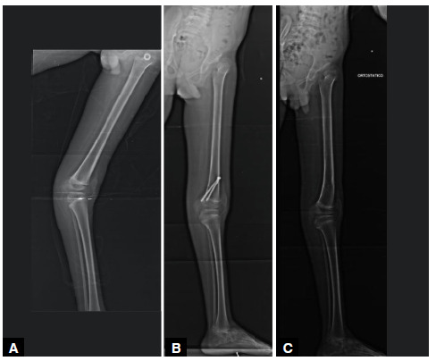
Introduction: The genu recurvatum is characterized by a hyperextension deformity of the knee in the sagittal plane. Among its causes are conditions such as arthrogryposis, cerebral palsy, poliomyelitis, sequelae of tibial tuberosity fracture and some syndromes with generalized joint hypermobility. Treatment of this deformity can be challenging and, to date, aggressive methods such as femur or tibial osteotomies are the most used for its correction.
Objective: This study aimed to describe a new surgical technique for correcting genu recurvatum.
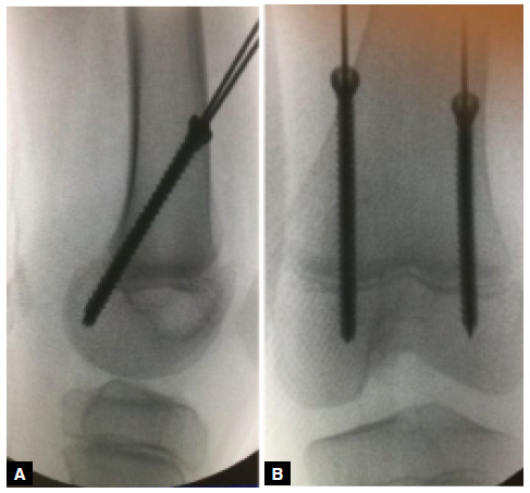
Methods: This is a prospective clinical study of children who underwent posterior hemiepiphysiodesis of the distal femur with transphyseal screws.
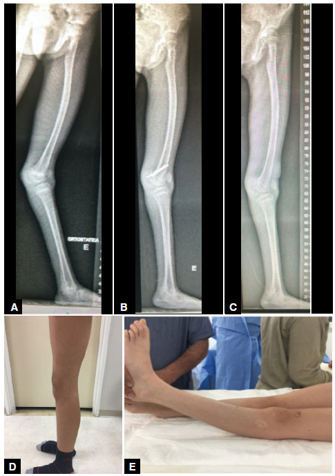
Results: The approach proved to be safe and useful for genu recurvatum deformities, with femoral or articular apex.
Conclusion: This approach shows great potential for correcting genu recurvatum in the developing skeleton, being an excellent alternative to the more aggressive methods currently used to treat this deformity. Level of evidence IV, Case Series.
期刊介绍:
A Revista Acta Ortopédica Brasileira, órgão oficial do Departamento de Ortopedia e Traumatologia da Faculdade de Medicina da Universidade de São Paulo (DOT/FMUSP), é publicada bimestralmente em seis edições ao ano (jan/fev, mar/abr, maio/jun, jul/ago, set/out e nov/dez) com versão em inglês disponível nos principais indexadores nacionais e internacionais e instituições de ensino do Brasil. Sendo hoje reconhecidamente uma importante contribuição para os especialistas da área com sua seriedade e árduo trabalho para as indexações já conquistadas.


 分享
分享
 求助内容:
求助内容: 应助结果提醒方式:
 扫码关注我们
