Kishin Tokuyama, Yusuke Inoue, Keiji Matsunaga, Yasunori Hamaguchi, Saori Sekimoto
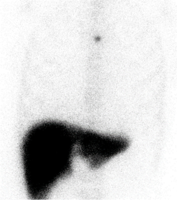
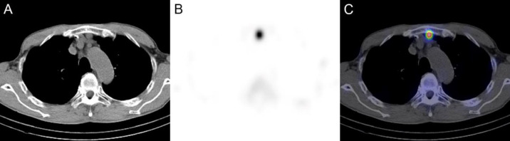
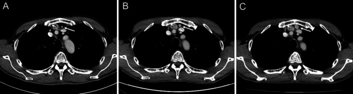
{"title":"<sup>99m</sup>Tc-Sn-colloid SPECT/CT in thoracic splenosis after esophageal cancer surgery.","authors":"Kishin Tokuyama, Yusuke Inoue, Keiji Matsunaga, Yasunori Hamaguchi, Saori Sekimoto","doi":"10.22038/AOJNMB.2023.73907.1515","DOIUrl":null,"url":null,"abstract":"<p><p>Splenosis occurs as a result of autotransplantation of splenic tissue following splenic injury or splenectomy. A 56-year-old man with esophageal cancer underwent thoracoscopic-assisted subtotal esophagectomy accompanied by three-field lymph node dissection, and retrosternal gastric tube reconstruction. The spleen was injured during the surgery and was removed. A retrosternal nodule of 12 mm in diameter was detected near the reconstructed gastric tube on computed tomography (CT) performed 3 years and 6 months postoperatively. Retrospectively, the nodule was observed in the same area on early postoperative CT and gradually increased in size. No accessory spleen was identified on the preoperative CT. Splenosis was suspected, and <sup>99m</sup>Tc-Sn-colloid single photon emission computed tomography (SPECT)/CT was performed. It revealed intense uptake in the retrosternal nodule, consistent with the diagnosis of thoracic splenosis. Subsequently, the patient has been under observation without treatment. <sup>99m</sup>Tc-labeled colloid SPECT/CT allowed confident diagnosis of thoracic splenosis following esophageal cancer surgery. This examination is considered valuable for the evaluation of ectopic splenic tissue.</p>","PeriodicalId":8503,"journal":{"name":"Asia Oceania Journal of Nuclear Medicine and Biology","volume":"12 1","pages":"61-64"},"PeriodicalIF":0.0000,"publicationDate":"2024-01-01","publicationTypes":"Journal Article","fieldsOfStudy":null,"isOpenAccess":false,"openAccessPdf":"https://www.ncbi.nlm.nih.gov/pmc/articles/PMC10757057/pdf/","citationCount":"0","resultStr":null,"platform":"Semanticscholar","paperid":null,"PeriodicalName":"Asia Oceania Journal of Nuclear Medicine and Biology","FirstCategoryId":"1085","ListUrlMain":"https://doi.org/10.22038/AOJNMB.2023.73907.1515","RegionNum":0,"RegionCategory":null,"ArticlePicture":[],"TitleCN":null,"AbstractTextCN":null,"PMCID":null,"EPubDate":"","PubModel":"","JCR":"Q3","JCRName":"Medicine","Score":null,"Total":0}
引用次数: 0
Abstract
Splenosis occurs as a result of autotransplantation of splenic tissue following splenic injury or splenectomy. A 56-year-old man with esophageal cancer underwent thoracoscopic-assisted subtotal esophagectomy accompanied by three-field lymph node dissection, and retrosternal gastric tube reconstruction. The spleen was injured during the surgery and was removed. A retrosternal nodule of 12 mm in diameter was detected near the reconstructed gastric tube on computed tomography (CT) performed 3 years and 6 months postoperatively. Retrospectively, the nodule was observed in the same area on early postoperative CT and gradually increased in size. No accessory spleen was identified on the preoperative CT. Splenosis was suspected, and 99mTc-Sn-colloid single photon emission computed tomography (SPECT)/CT was performed. It revealed intense uptake in the retrosternal nodule, consistent with the diagnosis of thoracic splenosis. Subsequently, the patient has been under observation without treatment. 99mTc-labeled colloid SPECT/CT allowed confident diagnosis of thoracic splenosis following esophageal cancer surgery. This examination is considered valuable for the evaluation of ectopic splenic tissue.


 分享
分享
 求助内容:
求助内容: 应助结果提醒方式:
 扫码关注我们
