{"title":"Physiological myocardial <sup>18</sup>F-FDG uptake pattern in oncologic PET/CT: comparison with findings in cardiac sarcoidosis.","authors":"Takashi Norikane, Yuka Yamamoto, Yasukage Takami, Katsuya Mitamura, Takuya Kobata, Yukito Maeda, Takahisa Noma, Yoshihiro Nishiyama","doi":"10.22038/AOJNMB.2023.70254.1490","DOIUrl":null,"url":null,"abstract":"<p><strong>Objectives: </strong>Physiological myocardial <sup>18</sup>F-fluorodeoxyglucose (<sup>18</sup>F-FDG) uptake in oncologic positron emission tomography (PET)/computed tomography (CT) is commonly observed with multiple variations under clinical fasting conditions. The purpose of the present study was to evaluate physiological myocardial <sup>18</sup>F-FDG uptake pattern by comparing with the results in cardiac sarcoidosis.</p><p><strong>Methods: </strong>A total of 174 examinations in 174 patients without cardiac disease and 27 examinations in 17 patients with cardiac sarcoidosis were performed. The polar map images generated from <sup>18</sup>F-FDG PET/CT data were visually assessed as \"basal-ring,\" \"focal,\" and \"focal on diffuse\" patterns. Semi-quantitative analysis was also performed using the regional relative <sup>18</sup>F-FDG uptake (% uptake).</p><p><strong>Results: </strong>On visual analysis, the \"focal on diffuse\" pattern was the most common in both examinations (43% and 59%, respectively). The physiological % uptake in the lateral and basal septal walls tended to be higher. Subgroup analysis showed significantly higher uptake in the mid-wall and left circumflex territory. In cardiac sarcoidosis patients, there was a significant difference only between segments 2 and 15 (p=0.04). No significant differences were observed between the base-mid-apical territory and coronary artery branch territory.</p><p><strong>Conclusion: </strong>High <sup>18</sup>F-FDG uptake in the basal septal walls is likely to be observed as both physiological uptake in patients without cardiac disease and pathological uptake in patients with cardiac sarcoidosis.</p>","PeriodicalId":8503,"journal":{"name":"Asia Oceania Journal of Nuclear Medicine and Biology","volume":"12 1","pages":"1-10"},"PeriodicalIF":0.0000,"publicationDate":"2024-01-01","publicationTypes":"Journal Article","fieldsOfStudy":null,"isOpenAccess":false,"openAccessPdf":"https://www.ncbi.nlm.nih.gov/pmc/articles/PMC10757061/pdf/","citationCount":"0","resultStr":null,"platform":"Semanticscholar","paperid":null,"PeriodicalName":"Asia Oceania Journal of Nuclear Medicine and Biology","FirstCategoryId":"1085","ListUrlMain":"https://doi.org/10.22038/AOJNMB.2023.70254.1490","RegionNum":0,"RegionCategory":null,"ArticlePicture":[],"TitleCN":null,"AbstractTextCN":null,"PMCID":null,"EPubDate":"","PubModel":"","JCR":"Q3","JCRName":"Medicine","Score":null,"Total":0}
引用次数: 0
Abstract
Objectives: Physiological myocardial 18F-fluorodeoxyglucose (18F-FDG) uptake in oncologic positron emission tomography (PET)/computed tomography (CT) is commonly observed with multiple variations under clinical fasting conditions. The purpose of the present study was to evaluate physiological myocardial 18F-FDG uptake pattern by comparing with the results in cardiac sarcoidosis.
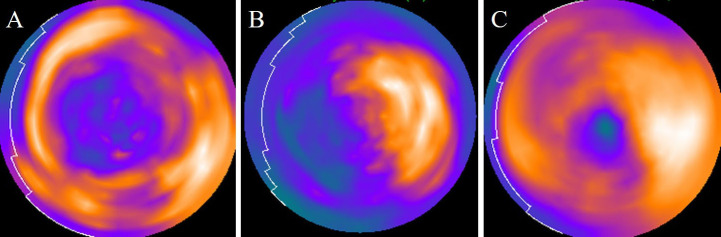
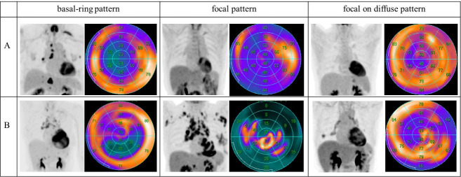
Methods: A total of 174 examinations in 174 patients without cardiac disease and 27 examinations in 17 patients with cardiac sarcoidosis were performed. The polar map images generated from 18F-FDG PET/CT data were visually assessed as "basal-ring," "focal," and "focal on diffuse" patterns. Semi-quantitative analysis was also performed using the regional relative 18F-FDG uptake (% uptake).
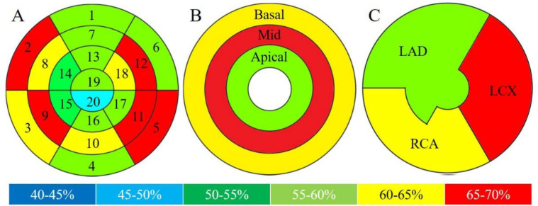
Results: On visual analysis, the "focal on diffuse" pattern was the most common in both examinations (43% and 59%, respectively). The physiological % uptake in the lateral and basal septal walls tended to be higher. Subgroup analysis showed significantly higher uptake in the mid-wall and left circumflex territory. In cardiac sarcoidosis patients, there was a significant difference only between segments 2 and 15 (p=0.04). No significant differences were observed between the base-mid-apical territory and coronary artery branch territory.
Conclusion: High 18F-FDG uptake in the basal septal walls is likely to be observed as both physiological uptake in patients without cardiac disease and pathological uptake in patients with cardiac sarcoidosis.


 分享
分享
 求助内容:
求助内容: 应助结果提醒方式:
 扫码关注我们
