Anwar Rahali, El Mehdi Aboulfath, Noureddine Njoumi, Mohammed Rebbani, Yasser El Brahmi, Mohammed Elfahssi, Abderrahman Elhjouji, Aziz Zentar, Abdelmounaim Ait Ali
{"title":"Sclerosing Encapsulating Peritonitis: Solving the Diagnosis Challenge of a Rare Entity.","authors":"Anwar Rahali, El Mehdi Aboulfath, Noureddine Njoumi, Mohammed Rebbani, Yasser El Brahmi, Mohammed Elfahssi, Abderrahman Elhjouji, Aziz Zentar, Abdelmounaim Ait Ali","doi":"10.1155/2023/4022487","DOIUrl":null,"url":null,"abstract":"<p><p>Sclerosing encapsulating peritonitis (SEP) is an unusual fibroinflammatory disease of the peritoneum marked by the development of a fibrous membrane enveloping generally the small intestines. The knowledge around this subject is not completely understood. And the etiology can be either idiopathic or secondary to several diseases, treatments, and/or medications. We present a case of a 52-year-old man suffering from atypical clinical symptoms including recurrent abdominal ascites and intestinal obstruction. An abdominal computed tomography showed findings typical of SEP. Therefore, the patient benefited from exploratory laparotomy, which confirmed the diagnosis of idiopathic SEP. Postoperatively, he again had an episode of bowel obstruction, but this was controlled with steroids. Diagnosis of SEP is a real challenge to surgeons, gastroenterologists, and radiologists. And imagery is very helpful to make the diagnosis. Consequently, it is imperative that all hospital practitioners should distinguish between this lesion and other etiology of acute peritonitis.</p>","PeriodicalId":9600,"journal":{"name":"Case Reports in Surgery","volume":"2023 ","pages":"4022487"},"PeriodicalIF":0.5000,"publicationDate":"2023-12-27","publicationTypes":"Journal Article","fieldsOfStudy":null,"isOpenAccess":false,"openAccessPdf":"https://www.ncbi.nlm.nih.gov/pmc/articles/PMC10764648/pdf/","citationCount":"0","resultStr":null,"platform":"Semanticscholar","paperid":null,"PeriodicalName":"Case Reports in Surgery","FirstCategoryId":"1085","ListUrlMain":"https://doi.org/10.1155/2023/4022487","RegionNum":0,"RegionCategory":null,"ArticlePicture":[],"TitleCN":null,"AbstractTextCN":null,"PMCID":null,"EPubDate":"2023/1/1 0:00:00","PubModel":"eCollection","JCR":"Q4","JCRName":"SURGERY","Score":null,"Total":0}
引用次数: 0
Abstract
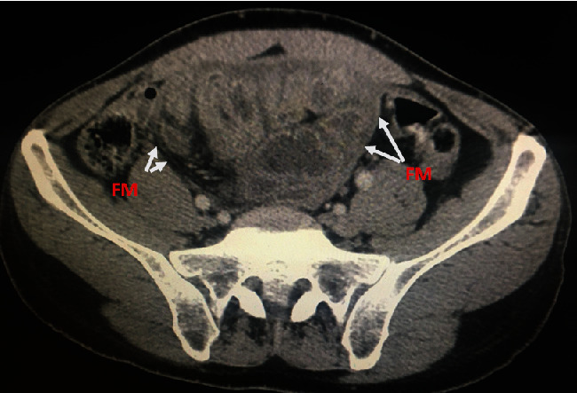
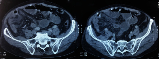
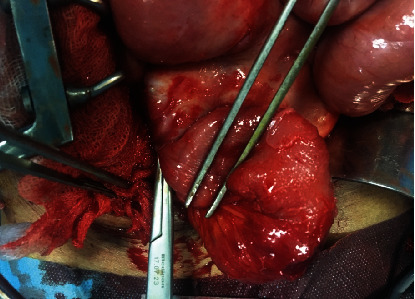
Sclerosing encapsulating peritonitis (SEP) is an unusual fibroinflammatory disease of the peritoneum marked by the development of a fibrous membrane enveloping generally the small intestines. The knowledge around this subject is not completely understood. And the etiology can be either idiopathic or secondary to several diseases, treatments, and/or medications. We present a case of a 52-year-old man suffering from atypical clinical symptoms including recurrent abdominal ascites and intestinal obstruction. An abdominal computed tomography showed findings typical of SEP. Therefore, the patient benefited from exploratory laparotomy, which confirmed the diagnosis of idiopathic SEP. Postoperatively, he again had an episode of bowel obstruction, but this was controlled with steroids. Diagnosis of SEP is a real challenge to surgeons, gastroenterologists, and radiologists. And imagery is very helpful to make the diagnosis. Consequently, it is imperative that all hospital practitioners should distinguish between this lesion and other etiology of acute peritonitis.


 分享
分享
 求助内容:
求助内容: 应助结果提醒方式:
 扫码关注我们
