Dan Guo, Jian Yang, Dianwei Liu, Pei Zhang, Hao Sun, Jingcheng Wang
{"title":"Human umbilical cord mesenchymal stem cells overexpressing RUNX1 promote tendon-bone healing by inhibiting osteolysis, enhancing osteogenesis and promoting angiogenesis.","authors":"Dan Guo, Jian Yang, Dianwei Liu, Pei Zhang, Hao Sun, Jingcheng Wang","doi":"10.1007/s13258-023-01478-3","DOIUrl":null,"url":null,"abstract":"<p><strong>Background: </strong>Rotator cuff injury (RCI) is a common shoulder injury, which is difficult to be completely repaired by surgery. Hence, new strategies are needed to promote the healing of tendon-bone.</p><p><strong>Objective: </strong>We aimed to investigate the effect of human umbilical cord mesenchymal stem cells (hUC-MSCs) overexpressing RUNX1 on the tendon-bone healing after RCI, and to further explore its mechanism.</p><p><strong>Methods: </strong>Lentiviral vector was used to mediate the overexpression of RUNX1. RUNX1-overexpressed UCB-MSCs (referred to as MSC-RUNX1) were co-cultured with osteoclasts, and TRAP staining was performed to observe the formation of osteoclasts. Then MSC-RUNX1 was cultured in osteogenic differentiation medium, Alizarin red staining was conducted to detect osteogenic differentiation. The expression of markers of osteogenesis and osteoclast was detected by RT-qPCR. EA. hy926 cells were co-cultured with MSC-RUNX1. Transwell assay was used to detect the migration, and the expression of angiogenesis related-genes VEGF and TGF-β was detected by RT-qPCR. The rat rotator cuff reconstruction model was established and MSCs were injected at the tendon-bone junction. Biomechanical test and micro-CT scanning were performed, and HE, Masson and Alcian Blue staining were used for histological evaluation of tendon-bone healing. TUNEL and PCNA immunofluorescence (IF) staining were performed to evaluate apoptosis and proliferation at the tendon-bone healing site. The levels of TNF-α, IL-6 and IL-8 in serum were detected by ELISA. The expression of CD31 and Endomucin that related to angiogenesis was detected by IF. Safranin O-fast and TRAP/CD40L immunohistochemical staining were used to assess the levels of osteoclasts and osteoblasts at the tendon-bone healing site.</p><p><strong>Results: </strong>hUC-MSCs overexpressing RUNX1 inhibited osteoclast formation and promoted osteogenic differentiation. MSC-RUNX1 could promote the migration and tube formation of EA. hy926 cells, and up-regulate the levels of VEGF and TGF-β. Model mice treated with MSC-RUNX1 partially restored the biomechanical indexes. Treatment of MSC-RUNX1 obviously increased the bone density, accompanied by the formation of new bone. In vivo experiments showed that MSC-RUNX1 treatment could promote tendon-bone healing and inhibit inflammatory response in rats. MSC-RUNX1 treatment also promoted angiogenesis at the tendon-bone healing site, while inhibiting osteoclast formation and promoting osteogenic differentiation.</p><p><strong>Conclusion: </strong>hUC-MSCs overexpressing RUNX1 can inhibit the formation of osteoclasts and differentiation of osteoblasts, promote angiogenesis and inhibit inflammation, thereby promoting tendon-bone healing after RCI.</p>","PeriodicalId":12675,"journal":{"name":"Genes & genomics","volume":" ","pages":"461-473"},"PeriodicalIF":1.7000,"publicationDate":"2024-04-01","publicationTypes":"Journal Article","fieldsOfStudy":null,"isOpenAccess":false,"openAccessPdf":"","citationCount":"0","resultStr":null,"platform":"Semanticscholar","paperid":null,"PeriodicalName":"Genes & genomics","FirstCategoryId":"99","ListUrlMain":"https://doi.org/10.1007/s13258-023-01478-3","RegionNum":4,"RegionCategory":"生物学","ArticlePicture":[],"TitleCN":null,"AbstractTextCN":null,"PMCID":null,"EPubDate":"2024/1/5 0:00:00","PubModel":"Epub","JCR":"Q4","JCRName":"BIOCHEMISTRY & MOLECULAR BIOLOGY","Score":null,"Total":0}
引用次数: 0
Abstract
Background: Rotator cuff injury (RCI) is a common shoulder injury, which is difficult to be completely repaired by surgery. Hence, new strategies are needed to promote the healing of tendon-bone.
Objective: We aimed to investigate the effect of human umbilical cord mesenchymal stem cells (hUC-MSCs) overexpressing RUNX1 on the tendon-bone healing after RCI, and to further explore its mechanism.
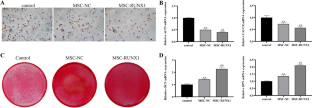
Methods: Lentiviral vector was used to mediate the overexpression of RUNX1. RUNX1-overexpressed UCB-MSCs (referred to as MSC-RUNX1) were co-cultured with osteoclasts, and TRAP staining was performed to observe the formation of osteoclasts. Then MSC-RUNX1 was cultured in osteogenic differentiation medium, Alizarin red staining was conducted to detect osteogenic differentiation. The expression of markers of osteogenesis and osteoclast was detected by RT-qPCR. EA. hy926 cells were co-cultured with MSC-RUNX1. Transwell assay was used to detect the migration, and the expression of angiogenesis related-genes VEGF and TGF-β was detected by RT-qPCR. The rat rotator cuff reconstruction model was established and MSCs were injected at the tendon-bone junction. Biomechanical test and micro-CT scanning were performed, and HE, Masson and Alcian Blue staining were used for histological evaluation of tendon-bone healing. TUNEL and PCNA immunofluorescence (IF) staining were performed to evaluate apoptosis and proliferation at the tendon-bone healing site. The levels of TNF-α, IL-6 and IL-8 in serum were detected by ELISA. The expression of CD31 and Endomucin that related to angiogenesis was detected by IF. Safranin O-fast and TRAP/CD40L immunohistochemical staining were used to assess the levels of osteoclasts and osteoblasts at the tendon-bone healing site.
Results: hUC-MSCs overexpressing RUNX1 inhibited osteoclast formation and promoted osteogenic differentiation. MSC-RUNX1 could promote the migration and tube formation of EA. hy926 cells, and up-regulate the levels of VEGF and TGF-β. Model mice treated with MSC-RUNX1 partially restored the biomechanical indexes. Treatment of MSC-RUNX1 obviously increased the bone density, accompanied by the formation of new bone. In vivo experiments showed that MSC-RUNX1 treatment could promote tendon-bone healing and inhibit inflammatory response in rats. MSC-RUNX1 treatment also promoted angiogenesis at the tendon-bone healing site, while inhibiting osteoclast formation and promoting osteogenic differentiation.
Conclusion: hUC-MSCs overexpressing RUNX1 can inhibit the formation of osteoclasts and differentiation of osteoblasts, promote angiogenesis and inhibit inflammation, thereby promoting tendon-bone healing after RCI.
期刊介绍:
Genes & Genomics is an official journal of the Korean Genetics Society (http://kgenetics.or.kr/). Although it is an official publication of the Genetics Society of Korea, membership of the Society is not required for contributors. It is a peer-reviewed international journal publishing print (ISSN 1976-9571) and online version (E-ISSN 2092-9293). It covers all disciplines of genetics and genomics from prokaryotes to eukaryotes from fundamental heredity to molecular aspects. The articles can be reviews, research articles, and short communications.


 分享
分享
 求助内容:
求助内容: 应助结果提醒方式:
 扫码关注我们
