{"title":"Three-Dimensional Imaging Analysis of the Developmental Process of Posterior Meniscofemoral Ligaments in Rat Embryos.","authors":"Momoko Nagai-Tanima, Kanon Ishida, Aoi Ishikawa, Shigehito Yamada, Tetsuya Takakuwa, Tomoki Aoyama","doi":"10.1159/000536108","DOIUrl":null,"url":null,"abstract":"<p><strong>Introduction: </strong>The posterior meniscofemoral ligament (pMFL) of knee joint is a ligament that runs posterior to the posterior cruciate ligament and it is known that the height of the pMFL attachment site causes meniscus avulsion. Therefore, understanding the three-dimensional (3D) structure of the pMFL attachment site is essential to better understand the pathogenesis of meniscus disorders. However, the developmental process of pMFL has not been well investigated. The purpose of this study was to analyze pMFL development in rat knee joints using 3D reconstructed images produced from episcopic fluorescence image capture (EFIC) images and examine its relationship with other knee joint components.</p><p><strong>Methods: </strong>Knee joints of Wistar rat embryos between embryonic day (E) 16 and E21 were observed with HE-stained tissues. Serial EFIC images of the hind limbs of E17-E21 were, respectively, captured from which 3D images were reconstructed and the features of pMFL structure: length and angle were measured. Besides, the chronological volume changes and the volume ratio of the knee joint components compared to E17 were calculated to identify the differences in growth by components.</p><p><strong>Results: </strong>pMFL was observed from E17 and was attached to the medial femoral condyle and lateral meniscus at all developmental stages, as in mature rats. The lack of marked variation in the attachment site and angle of the pMFL with the developmental stage indicates that the pMFL and surrounding knee joint components developed while maintaining their positional relationship from the onset of development.</p><p><strong>Conclusion: </strong>Current results may support to congenital etiology of meniscus disorder.</p>","PeriodicalId":9717,"journal":{"name":"Cells Tissues Organs","volume":" ","pages":"357-367"},"PeriodicalIF":1.9000,"publicationDate":"2024-01-01","publicationTypes":"Journal Article","fieldsOfStudy":null,"isOpenAccess":false,"openAccessPdf":"https://www.ncbi.nlm.nih.gov/pmc/articles/PMC11446320/pdf/","citationCount":"0","resultStr":null,"platform":"Semanticscholar","paperid":null,"PeriodicalName":"Cells Tissues Organs","FirstCategoryId":"99","ListUrlMain":"https://doi.org/10.1159/000536108","RegionNum":4,"RegionCategory":"生物学","ArticlePicture":[],"TitleCN":null,"AbstractTextCN":null,"PMCID":null,"EPubDate":"2024/1/5 0:00:00","PubModel":"Epub","JCR":"Q1","JCRName":"ANATOMY & MORPHOLOGY","Score":null,"Total":0}
引用次数: 0
Abstract
Introduction: The posterior meniscofemoral ligament (pMFL) of knee joint is a ligament that runs posterior to the posterior cruciate ligament and it is known that the height of the pMFL attachment site causes meniscus avulsion. Therefore, understanding the three-dimensional (3D) structure of the pMFL attachment site is essential to better understand the pathogenesis of meniscus disorders. However, the developmental process of pMFL has not been well investigated. The purpose of this study was to analyze pMFL development in rat knee joints using 3D reconstructed images produced from episcopic fluorescence image capture (EFIC) images and examine its relationship with other knee joint components.
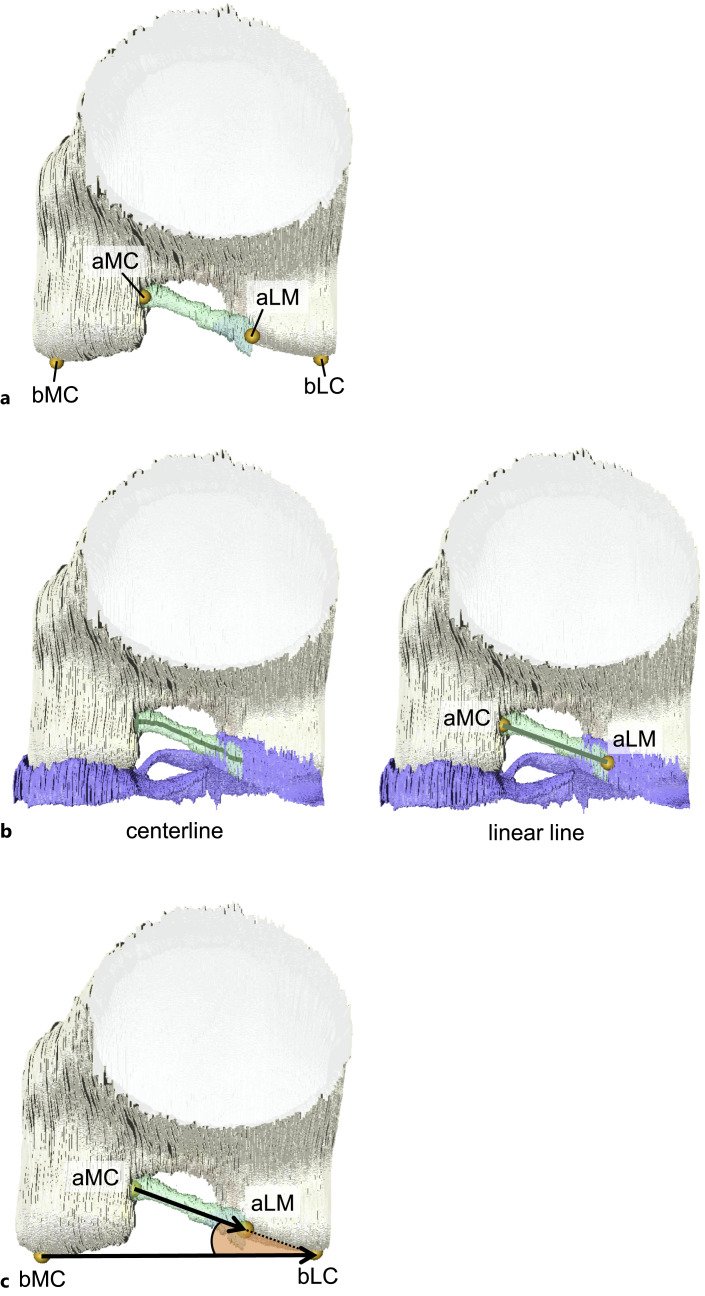
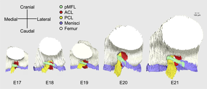
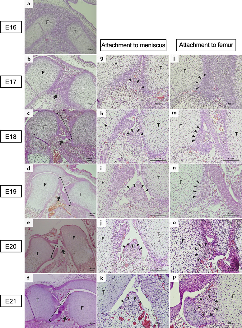
Methods: Knee joints of Wistar rat embryos between embryonic day (E) 16 and E21 were observed with HE-stained tissues. Serial EFIC images of the hind limbs of E17-E21 were, respectively, captured from which 3D images were reconstructed and the features of pMFL structure: length and angle were measured. Besides, the chronological volume changes and the volume ratio of the knee joint components compared to E17 were calculated to identify the differences in growth by components.
Results: pMFL was observed from E17 and was attached to the medial femoral condyle and lateral meniscus at all developmental stages, as in mature rats. The lack of marked variation in the attachment site and angle of the pMFL with the developmental stage indicates that the pMFL and surrounding knee joint components developed while maintaining their positional relationship from the onset of development.
Conclusion: Current results may support to congenital etiology of meniscus disorder.
期刊介绍:
''Cells Tissues Organs'' aims at bridging the gap between cell biology and developmental biology and the emerging fields of regenerative medicine (stem cell biology, tissue engineering, artificial organs, in vitro systems and transplantation biology). CTO offers a rapid and fair peer-review and exquisite reproduction quality. Special topic issues, entire issues of the journal devoted to a single research topic within the range of interests of the journal, are published at irregular intervals.


 分享
分享
 求助内容:
求助内容: 应助结果提醒方式:
 扫码关注我们
