{"title":"Dermoscopic Features of Heterotopic Salivary Gland Tissue in the Neck: Report of a Case.","authors":"Shinichiro Inoue, Takuya Maeda, Yuri Nagata, Teruki Yanagi, Hideyuki Ujiie","doi":"10.1159/000535740","DOIUrl":null,"url":null,"abstract":"<p><strong>Introduction: </strong>Heterotopic salivary gland tissue (HSGT) is diagnosed when salivary gland tissue is found in areas other than the major or minor salivary glands. No dermoscopic findings of HSGT have been reported.</p><p><strong>Case presentation: </strong>This case report gives the dermoscopic findings of a 45-year-old woman with HSGT who presented with a longstanding pale red macule on her neck, characterized by repeated clear fluid discharge. Dermoscopic examination showed a red dot with a pale pink halo and telangiectasia throughout the lesion. An excisional biopsy confirmed the diagnosis of HSGT, revealing the presence of mucous and serous salivary glandular structures within subcutaneous fat tissue. It has been reported that HSGT is associated with periductal lymphocytic infiltration, which was also observed in this case.</p><p><strong>Conclusion: </strong>We think that the \"red dot with a pale pink halo\" is a unique dermoscopic finding of HSGT, which is rarely observed in branchial anomalies.</p>","PeriodicalId":9619,"journal":{"name":"Case Reports in Dermatology","volume":"16 1","pages":"17-20"},"PeriodicalIF":0.8000,"publicationDate":"2024-01-09","publicationTypes":"Journal Article","fieldsOfStudy":null,"isOpenAccess":false,"openAccessPdf":"https://www.ncbi.nlm.nih.gov/pmc/articles/PMC10776130/pdf/","citationCount":"0","resultStr":null,"platform":"Semanticscholar","paperid":null,"PeriodicalName":"Case Reports in Dermatology","FirstCategoryId":"1085","ListUrlMain":"https://doi.org/10.1159/000535740","RegionNum":0,"RegionCategory":null,"ArticlePicture":[],"TitleCN":null,"AbstractTextCN":null,"PMCID":null,"EPubDate":"2024/1/1 0:00:00","PubModel":"eCollection","JCR":"Q4","JCRName":"DERMATOLOGY","Score":null,"Total":0}
引用次数: 0
Abstract
Introduction: Heterotopic salivary gland tissue (HSGT) is diagnosed when salivary gland tissue is found in areas other than the major or minor salivary glands. No dermoscopic findings of HSGT have been reported.
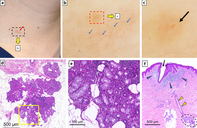
Case presentation: This case report gives the dermoscopic findings of a 45-year-old woman with HSGT who presented with a longstanding pale red macule on her neck, characterized by repeated clear fluid discharge. Dermoscopic examination showed a red dot with a pale pink halo and telangiectasia throughout the lesion. An excisional biopsy confirmed the diagnosis of HSGT, revealing the presence of mucous and serous salivary glandular structures within subcutaneous fat tissue. It has been reported that HSGT is associated with periductal lymphocytic infiltration, which was also observed in this case.
Conclusion: We think that the "red dot with a pale pink halo" is a unique dermoscopic finding of HSGT, which is rarely observed in branchial anomalies.


 分享
分享
 求助内容:
求助内容: 应助结果提醒方式:
 扫码关注我们
