Min-Sang Cha, Dae-Gon Kim, Yoon-Hyuk Huh, Lee-Ra Cho, Chan-Jin Park
{"title":"Three-dimensional morphometric study on the retromolar pad.","authors":"Min-Sang Cha, Dae-Gon Kim, Yoon-Hyuk Huh, Lee-Ra Cho, Chan-Jin Park","doi":"10.4047/jap.2023.15.6.302","DOIUrl":null,"url":null,"abstract":"<p><strong>Purpose: </strong>The aim of this study was to classify the shapes of retromolar pads and assess their morphometric differences using a 3D model.</p><p><strong>Materials and methods: </strong>Two hundred fully edentulous or Kennedy Class I partially edentulous patients (400 retromolar pads) were enrolled. Scan data of the definitive mandibular casts produced through functional impressions were obtained using a 3D laser scanner. Seven parameters (transverse diameter, longitudinal diameter, transverse-contour length, longitudinal-contour length, longitudinal/transverse diameter ratio, longitudinal/transverse-contour length ratio, and angle of the retromolar pad line to the residual alveolar ridge line) were measured using image analysis software. Subsequently, the pads were classified according to the shape. Statistical analyses were performed using 95% confidence intervals.</p><p><strong>Results: </strong>Classifying the retromolar pads into three shapes led to high intra-examiner reliability (Cronbach's alpha = 0.933). The pear shape was the most common (56.5%), followed by oval/round (27.7%) and triangular (15.8%) shapes. There were no significant differences between the left and right sides according to the shape and no significant differences in any parameter according to age. The transverse diameter and longitudinal/transverse diameter ratio differed between sexes (<i>P</i> < .05). The triangular shape had a significantly different transverse diameter, transverse-contour length, longitudinal/transverse diameter ratio, and longitudinal/transverse-contour length ratio compared with the pear and oval/round shapes (<i>P</i> < .05).</p><p><strong>Conclusion: </strong>From a clinical reliability standpoint, classifying retromolar pads into three shapes (oval/round, pear-shaped, and triangular) is effective. The differences in the sizes among the shapes were attributed to the transverse measurement values.</p>","PeriodicalId":51291,"journal":{"name":"Journal of Advanced Prosthodontics","volume":"15 6","pages":"302-314"},"PeriodicalIF":2.5000,"publicationDate":"2023-12-01","publicationTypes":"Journal Article","fieldsOfStudy":null,"isOpenAccess":false,"openAccessPdf":"https://www.ncbi.nlm.nih.gov/pmc/articles/PMC10774635/pdf/","citationCount":"0","resultStr":null,"platform":"Semanticscholar","paperid":null,"PeriodicalName":"Journal of Advanced Prosthodontics","FirstCategoryId":"3","ListUrlMain":"https://doi.org/10.4047/jap.2023.15.6.302","RegionNum":3,"RegionCategory":"医学","ArticlePicture":[],"TitleCN":null,"AbstractTextCN":null,"PMCID":null,"EPubDate":"2023/12/18 0:00:00","PubModel":"Epub","JCR":"Q1","JCRName":"DENTISTRY, ORAL SURGERY & MEDICINE","Score":null,"Total":0}
引用次数: 0
Abstract
Purpose: The aim of this study was to classify the shapes of retromolar pads and assess their morphometric differences using a 3D model.
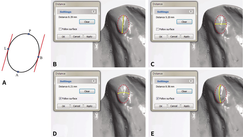
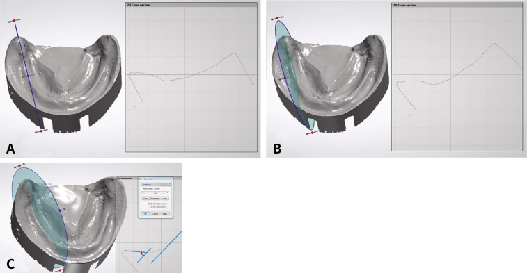
Materials and methods: Two hundred fully edentulous or Kennedy Class I partially edentulous patients (400 retromolar pads) were enrolled. Scan data of the definitive mandibular casts produced through functional impressions were obtained using a 3D laser scanner. Seven parameters (transverse diameter, longitudinal diameter, transverse-contour length, longitudinal-contour length, longitudinal/transverse diameter ratio, longitudinal/transverse-contour length ratio, and angle of the retromolar pad line to the residual alveolar ridge line) were measured using image analysis software. Subsequently, the pads were classified according to the shape. Statistical analyses were performed using 95% confidence intervals.
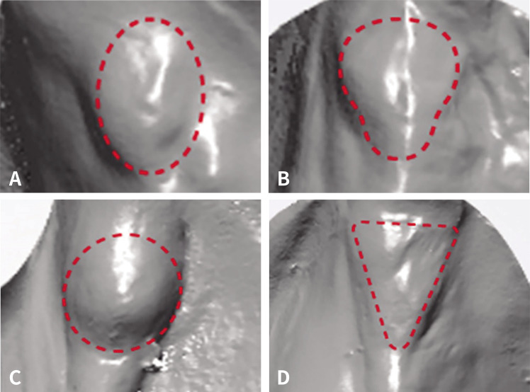
Results: Classifying the retromolar pads into three shapes led to high intra-examiner reliability (Cronbach's alpha = 0.933). The pear shape was the most common (56.5%), followed by oval/round (27.7%) and triangular (15.8%) shapes. There were no significant differences between the left and right sides according to the shape and no significant differences in any parameter according to age. The transverse diameter and longitudinal/transverse diameter ratio differed between sexes (P < .05). The triangular shape had a significantly different transverse diameter, transverse-contour length, longitudinal/transverse diameter ratio, and longitudinal/transverse-contour length ratio compared with the pear and oval/round shapes (P < .05).
Conclusion: From a clinical reliability standpoint, classifying retromolar pads into three shapes (oval/round, pear-shaped, and triangular) is effective. The differences in the sizes among the shapes were attributed to the transverse measurement values.
期刊介绍:
This journal aims to convey scientific and clinical progress in the field of prosthodontics and its related areas to many dental communities concerned with esthetic and functional restorations, occlusion, implants, prostheses, and biomaterials related to prosthodontics.
This journal publishes
• Original research data of high scientific merit in the field of diagnosis, function, esthetics and stomatognathic physiology related to prosthodontic rehabilitation, physiology and mechanics of occlusion, mechanical and biologic aspects of prosthodontic materials including dental implants.
• Review articles by experts on controversies and new developments in prosthodontics.
• Case reports if they provide or document new fundamental knowledge.


 分享
分享
 求助内容:
求助内容: 应助结果提醒方式:
 扫码关注我们
