Oxidative stress-induced temporal activation of ERK1/2 phosphorylates coreceptor of Wnt/β-catenin for myofibroblast formation in human lens epithelial cells.
IF 1.4 3区 医学Q4 BIOCHEMISTRY & MOLECULAR BIOLOGYMolecular VisionPub Date : 2023-10-20eCollection Date: 2023-01-01
Zaoxia Guo, Xiaopan Ma, Xi Chen, Rui Xue Zhang, Hong Yan
{"title":"Oxidative stress-induced temporal activation of ERK1/2 phosphorylates coreceptor of Wnt/β-catenin for myofibroblast formation in human lens epithelial cells.","authors":"Zaoxia Guo, Xiaopan Ma, Xi Chen, Rui Xue Zhang, Hong Yan","doi":"","DOIUrl":null,"url":null,"abstract":"<p><strong>Purpose: </strong>Posterior capsular opacification (PCO) is the most common complication postcataract surgery, and its underlying mechanisms involve epithelial-mesenchymal transition (EMT) of remnant lens epithelial cells (LECs) in response to drastic changes in stimuli in the intraocular environment, such as oxidative stress and growth factors. Wnt/β-catenin signaling is a major pathway mediating oxidative stress-induced EMT in LECs, but its interplay with other transduction pathways remains little known in the development of PCO. ERK1/2 signaling is the downstream component of a phosphorelay pathway in response to extracellular stimuli (e.g., reactive oxygen species), and its activation regulates multiple cellular processes, including proliferation and EMT. Thus, this study aimed to investigate how ERK1/2 signaling and Wnt/β-catenin pathway crosstalk in oxidative stress-induced EMT in LECs.</p><p><strong>Methods: </strong>Hydrogen peroxide (H<sub>2</sub>O<sub>2</sub>) at 50 μM treatment for 48 h was used to establish a moderate oxidative stress-induced EMT model in LECs. ERK1/2 signaling was inhibited using MEK1/2 inhibitor U0126 at 20 μM. Western blotting was used to quantify protein expression of various biomarkers of EMT and phosphorylated components in ERK1/2 and Wnt/β-catenin signaling. LEC proliferation was determined using an EdU staining assay and expression of proliferating cellular nuclear antigen (PCNA). Subcellular localization of biomarker proteins was visualized with immunofluorescent staining.</p><p><strong>Results: </strong>Under the moderate level of H<sub>2</sub>O<sub>2</sub>-induced EMT in LECs, ERK1/2 signaling was activated, as evidenced by a marked increase in the ratio of phosphorylated ERK1/2 to total ERK1/2 at early (i.e., 5-15 min) and late time points (i.e., 12 h); the canonical Wnt/β-catenin pathway was activated by H<sub>2</sub>O<sub>2</sub> at 48 h. LECs exposed to H<sub>2</sub>O<sub>2</sub> exhibited hyperproliferation and EMT; however, these were restored by inhibition of ERK1/2 signaling demonstrated by reduced DNA synthesis and PCNA expression for cellular proliferation and altered expression of various EMT protein markers, including E-cadherin, α-SMA, and vimentin. More importantly, inhibition of ERK1/2 signaling reduced β-catenin accumulation in the activated Wnt/β-catenin signaling cascade. Specifically, there was significant downregulation in the phosphorylation level of LRP6 at Ser 1490 and GSK-3β at Ser 9, the key coreceptor of Wnt and regulator of β-catenin, respectively.</p><p><strong>Conclusions: </strong>ERK1/2 signaling plays a crucial role in the moderate level of oxidative stress-induced EMT in LECs. Pharmacologically blocking ERK1/2 signaling significantly inhibited LEC proliferation and EMT. Mechanistically, ERK1/2 signaling regulated Wnt/β-catenin cascade by phosphorylating Wnt coreceptor LRP6 at Ser 1490 in the plasma membrane. These results shed light on a potential molecular switch of ERK1/2 and Wnt/β-catenin crosstalk underlying the development of PCO.</p>","PeriodicalId":18866,"journal":{"name":"Molecular Vision","volume":"29 ","pages":"206-216"},"PeriodicalIF":1.4000,"publicationDate":"2023-10-20","publicationTypes":"Journal Article","fieldsOfStudy":null,"isOpenAccess":false,"openAccessPdf":"https://www.ncbi.nlm.nih.gov/pmc/articles/PMC10784218/pdf/","citationCount":"0","resultStr":null,"platform":"Semanticscholar","paperid":null,"PeriodicalName":"Molecular Vision","FirstCategoryId":"3","ListUrlMain":"","RegionNum":3,"RegionCategory":"医学","ArticlePicture":[],"TitleCN":null,"AbstractTextCN":null,"PMCID":null,"EPubDate":"2023/1/1 0:00:00","PubModel":"eCollection","JCR":"Q4","JCRName":"BIOCHEMISTRY & MOLECULAR BIOLOGY","Score":null,"Total":0}
引用次数: 0
Abstract
Purpose: Posterior capsular opacification (PCO) is the most common complication postcataract surgery, and its underlying mechanisms involve epithelial-mesenchymal transition (EMT) of remnant lens epithelial cells (LECs) in response to drastic changes in stimuli in the intraocular environment, such as oxidative stress and growth factors. Wnt/β-catenin signaling is a major pathway mediating oxidative stress-induced EMT in LECs, but its interplay with other transduction pathways remains little known in the development of PCO. ERK1/2 signaling is the downstream component of a phosphorelay pathway in response to extracellular stimuli (e.g., reactive oxygen species), and its activation regulates multiple cellular processes, including proliferation and EMT. Thus, this study aimed to investigate how ERK1/2 signaling and Wnt/β-catenin pathway crosstalk in oxidative stress-induced EMT in LECs.
Methods: Hydrogen peroxide (H2O2) at 50 μM treatment for 48 h was used to establish a moderate oxidative stress-induced EMT model in LECs. ERK1/2 signaling was inhibited using MEK1/2 inhibitor U0126 at 20 μM. Western blotting was used to quantify protein expression of various biomarkers of EMT and phosphorylated components in ERK1/2 and Wnt/β-catenin signaling. LEC proliferation was determined using an EdU staining assay and expression of proliferating cellular nuclear antigen (PCNA). Subcellular localization of biomarker proteins was visualized with immunofluorescent staining.
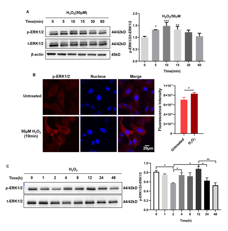
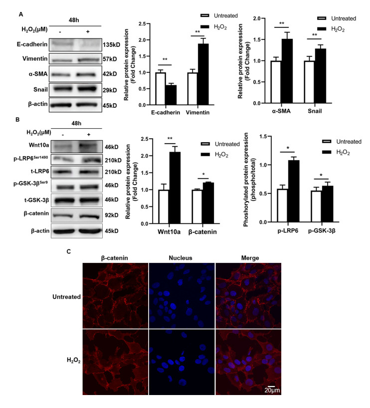
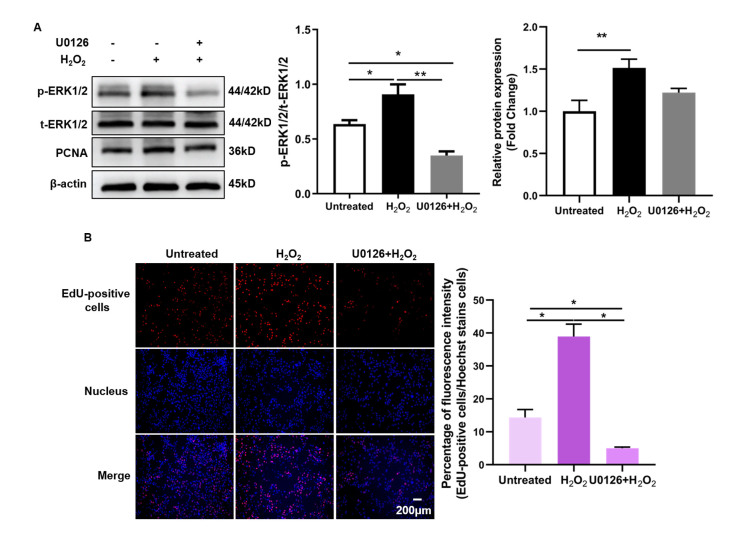
Results: Under the moderate level of H2O2-induced EMT in LECs, ERK1/2 signaling was activated, as evidenced by a marked increase in the ratio of phosphorylated ERK1/2 to total ERK1/2 at early (i.e., 5-15 min) and late time points (i.e., 12 h); the canonical Wnt/β-catenin pathway was activated by H2O2 at 48 h. LECs exposed to H2O2 exhibited hyperproliferation and EMT; however, these were restored by inhibition of ERK1/2 signaling demonstrated by reduced DNA synthesis and PCNA expression for cellular proliferation and altered expression of various EMT protein markers, including E-cadherin, α-SMA, and vimentin. More importantly, inhibition of ERK1/2 signaling reduced β-catenin accumulation in the activated Wnt/β-catenin signaling cascade. Specifically, there was significant downregulation in the phosphorylation level of LRP6 at Ser 1490 and GSK-3β at Ser 9, the key coreceptor of Wnt and regulator of β-catenin, respectively.
Conclusions: ERK1/2 signaling plays a crucial role in the moderate level of oxidative stress-induced EMT in LECs. Pharmacologically blocking ERK1/2 signaling significantly inhibited LEC proliferation and EMT. Mechanistically, ERK1/2 signaling regulated Wnt/β-catenin cascade by phosphorylating Wnt coreceptor LRP6 at Ser 1490 in the plasma membrane. These results shed light on a potential molecular switch of ERK1/2 and Wnt/β-catenin crosstalk underlying the development of PCO.
期刊介绍:
Molecular Vision is a peer-reviewed journal dedicated to the dissemination of research results in molecular biology, cell biology, and the genetics of the visual system (ocular and cortical).
Molecular Vision publishes articles presenting original research that has not previously been published and comprehensive articles reviewing the current status of a particular field or topic. Submissions to Molecular Vision are subjected to rigorous peer review. Molecular Vision does NOT publish preprints.
For authors, Molecular Vision provides a rapid means of communicating important results. Access to Molecular Vision is free and unrestricted, allowing the widest possible audience for your article. Digital publishing allows you to use color images freely (and without fees). Additionally, you may publish animations, sounds, or other supplementary information that clarifies or supports your article. Each of the authors of an article may also list an electronic mail address (which will be updated upon request) to give interested readers easy access to authors.
 分享
分享


 求助内容:
求助内容: 应助结果提醒方式:
 扫码关注我们
