Tevel Amiel, Oren Shauly, Ella Gilenson Istoyler, Michael Alterman, Naama Keshet, Sigal Mazor, Anna Pikovsky, Doron J. Aframian, Leo Joskowicz, Chen Nadler
{"title":"The anatomical pattern of ductal arborization in parotid glands using cone-beam computerized sialography","authors":"Tevel Amiel, Oren Shauly, Ella Gilenson Istoyler, Michael Alterman, Naama Keshet, Sigal Mazor, Anna Pikovsky, Doron J. Aframian, Leo Joskowicz, Chen Nadler","doi":"10.1002/ca.24136","DOIUrl":null,"url":null,"abstract":"<p>Cone-Beam Computed Tomography-Sialography (Sialo-CBCT) is used to demonstrate salivary ductal structure. This study aimed to conduct a volumetric analysis of the anatomical morphology of Normal-Appearing Glands (NAGs) in parotid sialo-CBCT. Our retrospective study included 14 parotid sialo-CBCT scans interpreted as NAGs in 11 patients with salivary gland impairment. The main duct length and width, as well as number and width of secondary and tertiary ducts were manually evaluated. We found that the main parotid duct showed an average width of 1.39 mm, 1.15 mm, and 0.98 mm, for the proximal, middle and distal thirds, respectively. The arborization pattern showed approximately 20% more tertiary (average number 11.1 ± 2.7) than secondary ducts (average number 9.0 ± 2.4) and approximately 8% narrower tertiary ducts (average width 0.65 ± 0.11 mm) compared to the secondary ducts (average width 0.77 ± 0.14 mm). Our anatomical analysis of NAGs in parotid sialo-CBCT demonstrated progressive narrowing of the main duct and increasing arborization and decreasing lumen size starting from the primary to the tertiary ducts. This is the most updated study regarding the anatomy of the parotid glands as demonstrated in sialo-CBCT. Our results may provide clinicians with the basic information for understanding aberration from normal morphology, as seen in salivary gland pathologies as well facilitate planning of treatment strategies, such as minimally invasive sialo-endoscopies, commonly practiced today.</p>","PeriodicalId":50687,"journal":{"name":"Clinical Anatomy","volume":"37 8","pages":"878-885"},"PeriodicalIF":2.3000,"publicationDate":"2024-01-25","publicationTypes":"Journal Article","fieldsOfStudy":null,"isOpenAccess":false,"openAccessPdf":"https://onlinelibrary.wiley.com/doi/epdf/10.1002/ca.24136","citationCount":"0","resultStr":null,"platform":"Semanticscholar","paperid":null,"PeriodicalName":"Clinical Anatomy","FirstCategoryId":"3","ListUrlMain":"https://onlinelibrary.wiley.com/doi/10.1002/ca.24136","RegionNum":4,"RegionCategory":"医学","ArticlePicture":[],"TitleCN":null,"AbstractTextCN":null,"PMCID":null,"EPubDate":"","PubModel":"","JCR":"Q1","JCRName":"ANATOMY & MORPHOLOGY","Score":null,"Total":0}
引用次数: 0
Abstract
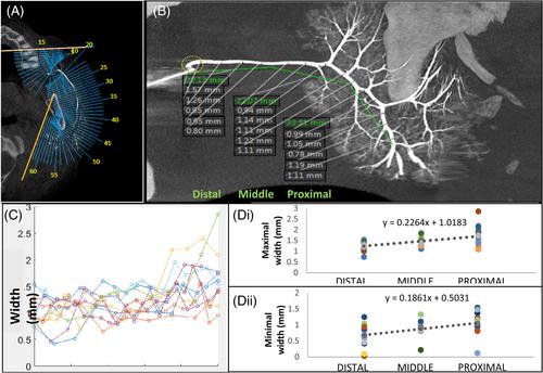
Cone-Beam Computed Tomography-Sialography (Sialo-CBCT) is used to demonstrate salivary ductal structure. This study aimed to conduct a volumetric analysis of the anatomical morphology of Normal-Appearing Glands (NAGs) in parotid sialo-CBCT. Our retrospective study included 14 parotid sialo-CBCT scans interpreted as NAGs in 11 patients with salivary gland impairment. The main duct length and width, as well as number and width of secondary and tertiary ducts were manually evaluated. We found that the main parotid duct showed an average width of 1.39 mm, 1.15 mm, and 0.98 mm, for the proximal, middle and distal thirds, respectively. The arborization pattern showed approximately 20% more tertiary (average number 11.1 ± 2.7) than secondary ducts (average number 9.0 ± 2.4) and approximately 8% narrower tertiary ducts (average width 0.65 ± 0.11 mm) compared to the secondary ducts (average width 0.77 ± 0.14 mm). Our anatomical analysis of NAGs in parotid sialo-CBCT demonstrated progressive narrowing of the main duct and increasing arborization and decreasing lumen size starting from the primary to the tertiary ducts. This is the most updated study regarding the anatomy of the parotid glands as demonstrated in sialo-CBCT. Our results may provide clinicians with the basic information for understanding aberration from normal morphology, as seen in salivary gland pathologies as well facilitate planning of treatment strategies, such as minimally invasive sialo-endoscopies, commonly practiced today.
期刊介绍:
Clinical Anatomy is the Official Journal of the American Association of Clinical Anatomists and the British Association of Clinical Anatomists. The goal of Clinical Anatomy is to provide a medium for the exchange of current information between anatomists and clinicians. This journal embraces anatomy in all its aspects as applied to medical practice. Furthermore, the journal assists physicians and other health care providers in keeping abreast of new methodologies for patient management and informs educators of new developments in clinical anatomy and teaching techniques. Clinical Anatomy publishes original and review articles of scientific, clinical, and educational interest. Papers covering the application of anatomic principles to the solution of clinical problems and/or the application of clinical observations to expand anatomic knowledge are welcomed.


 分享
分享
 求助内容:
求助内容: 应助结果提醒方式:
 扫码关注我们
