Anfisa Ayalon, Eran Greenbaum, Lily Okrent Smolar, Alexander Rubowitz
{"title":"Bilateral Subinternal Limiting Membrane Crystalline Deposits Secondary to Terson Syndrome.","authors":"Anfisa Ayalon, Eran Greenbaum, Lily Okrent Smolar, Alexander Rubowitz","doi":"10.1155/2024/8225960","DOIUrl":null,"url":null,"abstract":"<p><strong>Background: </strong>We report the case of bilateral, subinternal limiting membrane crystalline deposits in a patient with Terson syndrome, describe the possible pathogenesis, and highlight management. <i>Case Presentation</i>. A 24-year-old male with a history of traumatic massive parenchymal and subdural frontal hemorrhage presented to our clinic seven months after a motor vehicle accident, prolonged hospitalization, and rehabilitation, complaining of decreased vision in both eyes. The Snellen visual acuity was 1/60 in the right eye, and 6/60 in the left eye. Fundus examination showed an organized white vitreous hemorrhage in both eyes with almost no view of the retina. The anterior segments were normal. He underwent a 25-gauge pars plana vitrectomy in both eyes. During the surgery, golden crescent-shaped sediment consisting of small crystals was observed under the internal limiting membrane in both eyes: anterior to the inferior temporal vascular arcade in the right eye and posterior to it in the left eye. Internal limiting membrane (ILM) peeling after staining with ILM-blue dye was performed in the left eye, where the finding involved the macula. One year after the surgery, visual acuity significantly improved to 6/8.5 on the right and 6/6 on the left. Epiretinal membrane formation was observed in the right eye, where ILM peeling was not performed.</p><p><strong>Conclusion: </strong>Subinternal limiting membrane crystalline deposit finding is a rare condition. Consider performing internal limiting membrane peeling and sediment removal in cases with macular involvement. In cases where crystals are concentrated outside of the macula, follow-up may be considered.</p>","PeriodicalId":9603,"journal":{"name":"Case Reports in Ophthalmological Medicine","volume":"2024 ","pages":"8225960"},"PeriodicalIF":0.4000,"publicationDate":"2024-01-17","publicationTypes":"Journal Article","fieldsOfStudy":null,"isOpenAccess":false,"openAccessPdf":"https://www.ncbi.nlm.nih.gov/pmc/articles/PMC10807941/pdf/","citationCount":"0","resultStr":null,"platform":"Semanticscholar","paperid":null,"PeriodicalName":"Case Reports in Ophthalmological Medicine","FirstCategoryId":"1085","ListUrlMain":"https://doi.org/10.1155/2024/8225960","RegionNum":0,"RegionCategory":null,"ArticlePicture":[],"TitleCN":null,"AbstractTextCN":null,"PMCID":null,"EPubDate":"2024/1/1 0:00:00","PubModel":"eCollection","JCR":"Q4","JCRName":"OPHTHALMOLOGY","Score":null,"Total":0}
引用次数: 0
Abstract
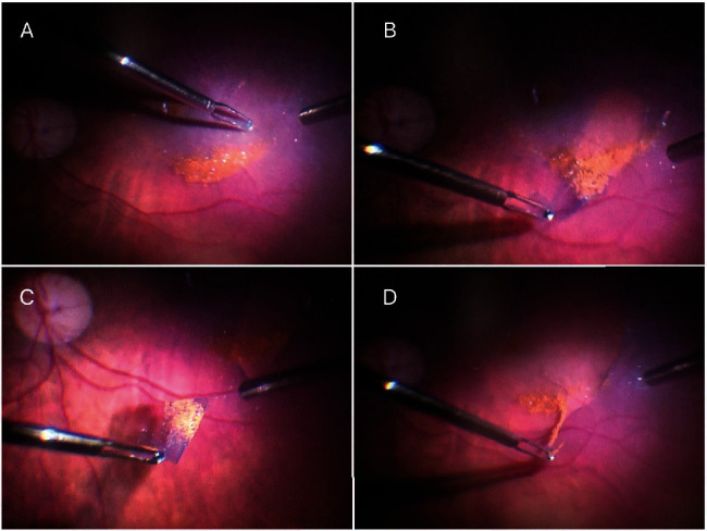
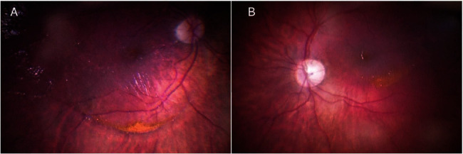
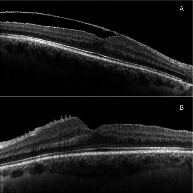
Background: We report the case of bilateral, subinternal limiting membrane crystalline deposits in a patient with Terson syndrome, describe the possible pathogenesis, and highlight management. Case Presentation. A 24-year-old male with a history of traumatic massive parenchymal and subdural frontal hemorrhage presented to our clinic seven months after a motor vehicle accident, prolonged hospitalization, and rehabilitation, complaining of decreased vision in both eyes. The Snellen visual acuity was 1/60 in the right eye, and 6/60 in the left eye. Fundus examination showed an organized white vitreous hemorrhage in both eyes with almost no view of the retina. The anterior segments were normal. He underwent a 25-gauge pars plana vitrectomy in both eyes. During the surgery, golden crescent-shaped sediment consisting of small crystals was observed under the internal limiting membrane in both eyes: anterior to the inferior temporal vascular arcade in the right eye and posterior to it in the left eye. Internal limiting membrane (ILM) peeling after staining with ILM-blue dye was performed in the left eye, where the finding involved the macula. One year after the surgery, visual acuity significantly improved to 6/8.5 on the right and 6/6 on the left. Epiretinal membrane formation was observed in the right eye, where ILM peeling was not performed.
Conclusion: Subinternal limiting membrane crystalline deposit finding is a rare condition. Consider performing internal limiting membrane peeling and sediment removal in cases with macular involvement. In cases where crystals are concentrated outside of the macula, follow-up may be considered.


 分享
分享
 求助内容:
求助内容: 应助结果提醒方式:
 扫码关注我们
