Laura Guerrero, Lorena Carmona-Rodríguez, Fátima Milhano Santos, Sergio Ciordia, Luiz Stark, Loreto Hierro, Pablo Pérez-Montero, David Vicent, Fernando J. Corrales
{"title":"Molecular basis of progressive familial intrahepatic cholestasis 3. A proteomics study","authors":"Laura Guerrero, Lorena Carmona-Rodríguez, Fátima Milhano Santos, Sergio Ciordia, Luiz Stark, Loreto Hierro, Pablo Pérez-Montero, David Vicent, Fernando J. Corrales","doi":"10.1002/biof.2041","DOIUrl":null,"url":null,"abstract":"<p>Progressive familial intrahepatic cholestasis type 3 (PFIC3) is a severe rare liver disease that affects between 1/50,000 and 1/100,000 children. In physiological conditions, bile is produced by the liver and stored in the gallbladder, and then it flows to the small intestine to play its role in fat digestion. To prevent tissue damage, bile acids (BAs) are kept in phospholipid micelles. Mutations in phosphatidyl choline transporter ABCB4 (<i>MDR3</i>) lead to intrahepatic accumulation of free BAs that result in liver damage. PFIC3 onset usually occurs at early ages, progresses rapidly, and the prognosis is poor. Currently, besides the palliative use of ursodeoxycholate, the only available treatment for this disease is liver transplantation, which is really challenging for short-aged patients. To gain insight into the pathogenesis of PFIC3 we have performed an integrated proteomics and phosphoproteomics study in human liver samples to then validate the emerging functional hypotheses in a PFIC3 murine model. We identified 6246 protein groups, 324 proteins among them showing differential expression between control and PFIC3. The phosphoproteomic analysis allowed the identification of 5090 phosphopeptides, from which 215 corresponding to 157 protein groups, were differentially phosphorylated in PFIC3, including MDR3. Regulation of essential cellular processes and structures, such as inflammation, metabolic reprogramming, cytoskeleton and extracellular matrix remodeling, and cell proliferation, were identified as the main drivers of the disease. Our results provide a strong molecular background that significantly contributes to a better understanding of PFIC3 and provides new concepts that might prove useful in the clinical management of patients.</p>","PeriodicalId":8923,"journal":{"name":"BioFactors","volume":"50 4","pages":"794-809"},"PeriodicalIF":5.0000,"publicationDate":"2024-01-29","publicationTypes":"Journal Article","fieldsOfStudy":null,"isOpenAccess":false,"openAccessPdf":"https://onlinelibrary.wiley.com/doi/epdf/10.1002/biof.2041","citationCount":"0","resultStr":null,"platform":"Semanticscholar","paperid":null,"PeriodicalName":"BioFactors","FirstCategoryId":"99","ListUrlMain":"https://iubmb.onlinelibrary.wiley.com/doi/10.1002/biof.2041","RegionNum":3,"RegionCategory":"生物学","ArticlePicture":[],"TitleCN":null,"AbstractTextCN":null,"PMCID":null,"EPubDate":"","PubModel":"","JCR":"Q1","JCRName":"BIOCHEMISTRY & MOLECULAR BIOLOGY","Score":null,"Total":0}
引用次数: 0
Abstract
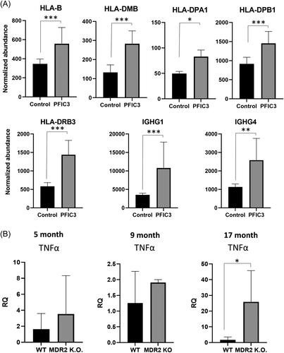
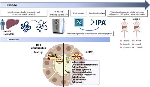
Progressive familial intrahepatic cholestasis type 3 (PFIC3) is a severe rare liver disease that affects between 1/50,000 and 1/100,000 children. In physiological conditions, bile is produced by the liver and stored in the gallbladder, and then it flows to the small intestine to play its role in fat digestion. To prevent tissue damage, bile acids (BAs) are kept in phospholipid micelles. Mutations in phosphatidyl choline transporter ABCB4 (MDR3) lead to intrahepatic accumulation of free BAs that result in liver damage. PFIC3 onset usually occurs at early ages, progresses rapidly, and the prognosis is poor. Currently, besides the palliative use of ursodeoxycholate, the only available treatment for this disease is liver transplantation, which is really challenging for short-aged patients. To gain insight into the pathogenesis of PFIC3 we have performed an integrated proteomics and phosphoproteomics study in human liver samples to then validate the emerging functional hypotheses in a PFIC3 murine model. We identified 6246 protein groups, 324 proteins among them showing differential expression between control and PFIC3. The phosphoproteomic analysis allowed the identification of 5090 phosphopeptides, from which 215 corresponding to 157 protein groups, were differentially phosphorylated in PFIC3, including MDR3. Regulation of essential cellular processes and structures, such as inflammation, metabolic reprogramming, cytoskeleton and extracellular matrix remodeling, and cell proliferation, were identified as the main drivers of the disease. Our results provide a strong molecular background that significantly contributes to a better understanding of PFIC3 and provides new concepts that might prove useful in the clinical management of patients.
期刊介绍:
BioFactors, a journal of the International Union of Biochemistry and Molecular Biology, is devoted to the rapid publication of highly significant original research articles and reviews in experimental biology in health and disease.
The word “biofactors” refers to the many compounds that regulate biological functions. Biological factors comprise many molecules produced or modified by living organisms, and present in many essential systems like the blood, the nervous or immunological systems. A non-exhaustive list of biological factors includes neurotransmitters, cytokines, chemokines, hormones, coagulation factors, transcription factors, signaling molecules, receptor ligands and many more. In the group of biofactors we can accommodate several classical molecules not synthetized in the body such as vitamins, micronutrients or essential trace elements.
In keeping with this unified view of biochemistry, BioFactors publishes research dealing with the identification of new substances and the elucidation of their functions at the biophysical, biochemical, cellular and human level as well as studies revealing novel functions of already known biofactors. The journal encourages the submission of studies that use biochemistry, biophysics, cell and molecular biology and/or cell signaling approaches.


 分享
分享
 求助内容:
求助内容: 应助结果提醒方式:
 扫码关注我们
