RNASeq Analysis for Accurate Identification of Fusion Partners in Tumor Specific Translocations Detected by Standard FISH Probes in Hematologic Malignancies.
Prasad Koduru, Weina Chen, Franklin Fuda, Gurbakhash Kaur, Farrukh Awan, Samuel John, Rolando Garcia, Jeffrey Gagan
{"title":"RNASeq Analysis for Accurate Identification of Fusion Partners in Tumor Specific Translocations Detected by Standard FISH Probes in Hematologic Malignancies.","authors":"Prasad Koduru, Weina Chen, Franklin Fuda, Gurbakhash Kaur, Farrukh Awan, Samuel John, Rolando Garcia, Jeffrey Gagan","doi":"10.1177/2632010X241230262","DOIUrl":null,"url":null,"abstract":"<p><strong>Background: </strong>Fluorescence labeled DNA probes and in situ hybridization methods had shorter turn round time for results revolutionized their clinical application. Signals obtained from these probes are highly specific, yet they can produce fusion signals not necessarily representing fusion of actual genes due to other genes included in the probe design. In this study we evaluated discordance between cytogenetic, FISH and RNAseq results in 3 different patients with hematologic malignancies and illustrated the need to perform next generation sequencing (NGS) or RNASeq to accurately interpret FISH results.</p><p><strong>Methods: </strong>Bone marrow or peripheral blood karyotypes and FISH were performed to detect recurring translocations associated with hematologic malignancies in clinical samples routinely referred to our clinical cytogenetics laboratory. When required, NGS was performed on DNA and RNA libraries to detect somatic alterations and gene fusions in some of these specimens. Discordance in results between these methods is further evaluated.</p><p><strong>Results: </strong>For a patient with plasma cell leukemia standard <i>FGFR3</i> / <i>IGH</i> dual fusion FISH assay detected fusion that was interpreted as <i>FGFR3</i>-positive leukemia, whereas NGS/RNASeq detected <i>NSD2::IGH</i>. For a pediatric acute lymphoblastic leukemia patient, a genetic diagnosis of <i>PDGFRB</i>-positive ALL was rendered because the <i>PDGFRB</i> break-apart probe detected clonal rearrangement, whereas NGS detected <i>MEF2D::CSF1R</i>. A <i>MYC</i>-positive B-prolymphocytic leukemia was rendered for another patient with a cytogenetically identified t(8;14) and <i>MYC::IGH</i> by FISH, whereas NGS detected a novel <i>PVT1::RCOR1</i> not previously reported.</p><p><strong>Conclusions: </strong>These are 3 cases in a series of several other concordant results, nevertheless, elucidate limitations when interpreting FISH results in clinical applications, particularly when other genes are included in probe design. In addition, when the observed FISH signals are atypical, this study illustrates the necessity to perform complementary laboratory assays, such as NGS and/or RNASeq, to accurately identify fusion genes in tumorigenic translocations.</p>","PeriodicalId":53204,"journal":{"name":"Clinical Pathology","volume":"17 ","pages":"2632010X241230262"},"PeriodicalIF":1.9000,"publicationDate":"2024-02-16","publicationTypes":"Journal Article","fieldsOfStudy":null,"isOpenAccess":false,"openAccessPdf":"https://www.ncbi.nlm.nih.gov/pmc/articles/PMC10874141/pdf/","citationCount":"0","resultStr":null,"platform":"Semanticscholar","paperid":null,"PeriodicalName":"Clinical Pathology","FirstCategoryId":"1085","ListUrlMain":"https://doi.org/10.1177/2632010X241230262","RegionNum":0,"RegionCategory":null,"ArticlePicture":[],"TitleCN":null,"AbstractTextCN":null,"PMCID":null,"EPubDate":"2024/1/1 0:00:00","PubModel":"eCollection","JCR":"Q3","JCRName":"PATHOLOGY","Score":null,"Total":0}
引用次数: 0
Abstract
Background: Fluorescence labeled DNA probes and in situ hybridization methods had shorter turn round time for results revolutionized their clinical application. Signals obtained from these probes are highly specific, yet they can produce fusion signals not necessarily representing fusion of actual genes due to other genes included in the probe design. In this study we evaluated discordance between cytogenetic, FISH and RNAseq results in 3 different patients with hematologic malignancies and illustrated the need to perform next generation sequencing (NGS) or RNASeq to accurately interpret FISH results.
Methods: Bone marrow or peripheral blood karyotypes and FISH were performed to detect recurring translocations associated with hematologic malignancies in clinical samples routinely referred to our clinical cytogenetics laboratory. When required, NGS was performed on DNA and RNA libraries to detect somatic alterations and gene fusions in some of these specimens. Discordance in results between these methods is further evaluated.
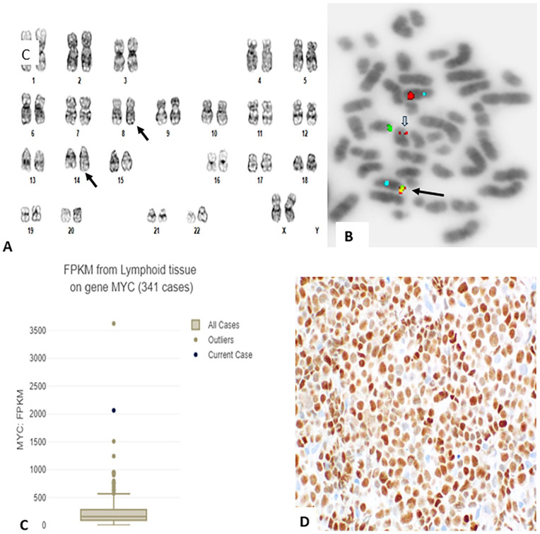
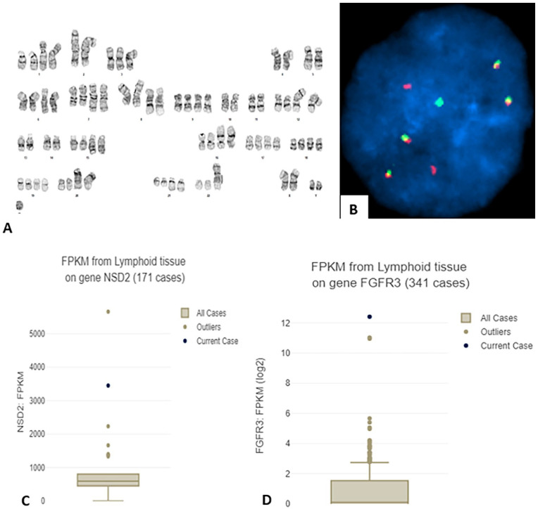
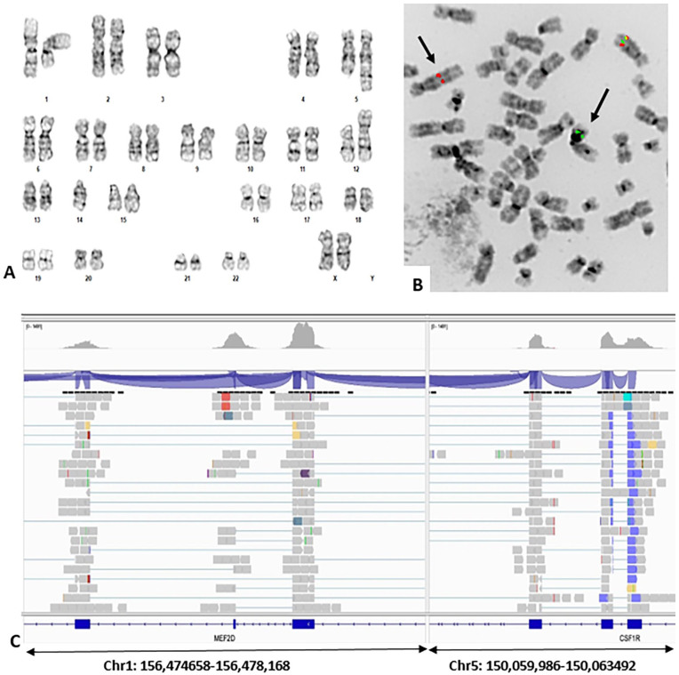
Results: For a patient with plasma cell leukemia standard FGFR3 / IGH dual fusion FISH assay detected fusion that was interpreted as FGFR3-positive leukemia, whereas NGS/RNASeq detected NSD2::IGH. For a pediatric acute lymphoblastic leukemia patient, a genetic diagnosis of PDGFRB-positive ALL was rendered because the PDGFRB break-apart probe detected clonal rearrangement, whereas NGS detected MEF2D::CSF1R. A MYC-positive B-prolymphocytic leukemia was rendered for another patient with a cytogenetically identified t(8;14) and MYC::IGH by FISH, whereas NGS detected a novel PVT1::RCOR1 not previously reported.
Conclusions: These are 3 cases in a series of several other concordant results, nevertheless, elucidate limitations when interpreting FISH results in clinical applications, particularly when other genes are included in probe design. In addition, when the observed FISH signals are atypical, this study illustrates the necessity to perform complementary laboratory assays, such as NGS and/or RNASeq, to accurately identify fusion genes in tumorigenic translocations.


 分享
分享
 求助内容:
求助内容: 应助结果提醒方式:
 扫码关注我们
