Waldemar Leszczyński, Wojciech Kazimierczak, Adam Lemanowicz, Zbigniew Serafin
{"title":"Texture analysis of chest X-ray images for the diagnosis of COVID-19 pneumonia.","authors":"Waldemar Leszczyński, Wojciech Kazimierczak, Adam Lemanowicz, Zbigniew Serafin","doi":"10.5114/pjr.2024.134818","DOIUrl":null,"url":null,"abstract":"<p><strong>Purpose: </strong>Medical imaging is one of the main methods of diagnosing COVID-19, along with real-time reverse trans-cription-polymerase chain reaction (RT-PCR) tests. The purpose of the study was to analyse the texture parameters of chest X-rays (CXR) of patients suspected of having COVID-19.</p><p><strong>Material and methods: </strong>Texture parameters of the CXRs of 70 patients with symptoms typical of COVID-19 infection were analysed using LIFEx software. The regions of interest (ROIs) included each lung separately, for which 57 para-meters were tested. The control group consisted of 30 healthy, age-matched patients with no pathological findings in CXRs.</p><p><strong>Results: </strong>According to the ROC analysis, 13 of the tested parameters differentiate the radiological image of lungs with COVID-19 features from the image of healthy lungs: GLRLM_LRHGE (AUC 0.91); DISCRETIZED_Q3 (AUC 0.90); GLZLM_HGZE (AUC 0.90); GLRLM_HGRE (AUC 0.89); DISCRETIZED_mean (AUC 0.89); DISCRETIZED_Q2 (AUC 0.61); GLRLM_SRHGE (AUC 0.87); GLZLM_LZHGE (AUC 0.87); GLZLM_SZHGE (AUC 0.84); DISCRETIZED_Q1 (AUC 0.81); NGLDM_Coarseness (AUC 0.70); DISCRETIZED_std (AUC 0.64); CONVENTIONAL_Q2 (AUC 0.61).</p><p><strong>Conclusions: </strong>Selected texture parameters of radiological CXRs make it possible to distinguish COVID-19 features from healthy ones.</p>","PeriodicalId":94174,"journal":{"name":"Polish journal of radiology","volume":"89 ","pages":"e49-e53"},"PeriodicalIF":0.0000,"publicationDate":"2024-01-25","publicationTypes":"Journal Article","fieldsOfStudy":null,"isOpenAccess":false,"openAccessPdf":"https://www.ncbi.nlm.nih.gov/pmc/articles/PMC10867972/pdf/","citationCount":"0","resultStr":null,"platform":"Semanticscholar","paperid":null,"PeriodicalName":"Polish journal of radiology","FirstCategoryId":"1085","ListUrlMain":"https://doi.org/10.5114/pjr.2024.134818","RegionNum":0,"RegionCategory":null,"ArticlePicture":[],"TitleCN":null,"AbstractTextCN":null,"PMCID":null,"EPubDate":"2024/1/1 0:00:00","PubModel":"eCollection","JCR":"","JCRName":"","Score":null,"Total":0}
引用次数: 0
Abstract
Purpose: Medical imaging is one of the main methods of diagnosing COVID-19, along with real-time reverse trans-cription-polymerase chain reaction (RT-PCR) tests. The purpose of the study was to analyse the texture parameters of chest X-rays (CXR) of patients suspected of having COVID-19.
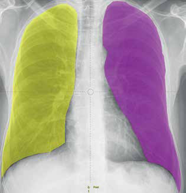
Material and methods: Texture parameters of the CXRs of 70 patients with symptoms typical of COVID-19 infection were analysed using LIFEx software. The regions of interest (ROIs) included each lung separately, for which 57 para-meters were tested. The control group consisted of 30 healthy, age-matched patients with no pathological findings in CXRs.
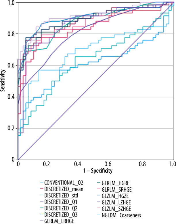
Results: According to the ROC analysis, 13 of the tested parameters differentiate the radiological image of lungs with COVID-19 features from the image of healthy lungs: GLRLM_LRHGE (AUC 0.91); DISCRETIZED_Q3 (AUC 0.90); GLZLM_HGZE (AUC 0.90); GLRLM_HGRE (AUC 0.89); DISCRETIZED_mean (AUC 0.89); DISCRETIZED_Q2 (AUC 0.61); GLRLM_SRHGE (AUC 0.87); GLZLM_LZHGE (AUC 0.87); GLZLM_SZHGE (AUC 0.84); DISCRETIZED_Q1 (AUC 0.81); NGLDM_Coarseness (AUC 0.70); DISCRETIZED_std (AUC 0.64); CONVENTIONAL_Q2 (AUC 0.61).
Conclusions: Selected texture parameters of radiological CXRs make it possible to distinguish COVID-19 features from healthy ones.


 分享
分享

 求助内容:
求助内容: 应助结果提醒方式:
 扫码关注我们
