{"title":"Impact of Stem Cells on Reparative Regeneration in Abdominal and Dorsal Skin in the Rat.","authors":"Evgeniya Kananykhina, Andrey Elchaninov, Galina Bolshakova","doi":"10.3390/jdb12010006","DOIUrl":null,"url":null,"abstract":"<p><p>A characteristic feature of repair processes in mammals is the formation of scar tissue at the site of injury, which is designed to quickly prevent contact between the internal environment of the organism and the external environment. Despite this general pattern, different organs differ in the degree of severity of scar changes in response to injury. One of the areas in which regeneration after wounding leads to the formation of a structure close to the original one is the abdominal skin of laboratory rats. Finding out the reasons for such a phenomenon is essential for the development of ways to stimulate full regeneration. The model of skin wound healing in the abdominal region of laboratory animals was reproduced in this work. It was found that the wound surface is completely epithelialized on the abdomen by 20 days, while on the back-by 30 days. The qPCR method revealed higher expression of marker genes of skin stem cells (<i>Sox9</i>, <i>Lgr6</i>, <i>Gli1</i>, <i>Lrig1</i>) in the intact skin of the abdomen compared to the back, which corresponded to a greater number of hairs with which stem cells are associated on the abdomen compared to the back. Considering that some stem cell populations are associated with hair, it can be suggested that one of the factors in faster regeneration of abdominal skin in the rat is the greater number of stem cells in this area.</p>","PeriodicalId":15563,"journal":{"name":"Journal of Developmental Biology","volume":"12 1","pages":""},"PeriodicalIF":2.5000,"publicationDate":"2024-01-27","publicationTypes":"Journal Article","fieldsOfStudy":null,"isOpenAccess":false,"openAccessPdf":"https://www.ncbi.nlm.nih.gov/pmc/articles/PMC10885081/pdf/","citationCount":"0","resultStr":null,"platform":"Semanticscholar","paperid":null,"PeriodicalName":"Journal of Developmental Biology","FirstCategoryId":"1085","ListUrlMain":"https://doi.org/10.3390/jdb12010006","RegionNum":0,"RegionCategory":null,"ArticlePicture":[],"TitleCN":null,"AbstractTextCN":null,"PMCID":null,"EPubDate":"","PubModel":"","JCR":"Q3","JCRName":"DEVELOPMENTAL BIOLOGY","Score":null,"Total":0}
引用次数: 0
Abstract
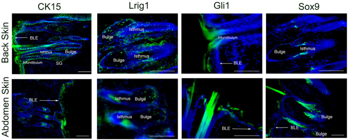
A characteristic feature of repair processes in mammals is the formation of scar tissue at the site of injury, which is designed to quickly prevent contact between the internal environment of the organism and the external environment. Despite this general pattern, different organs differ in the degree of severity of scar changes in response to injury. One of the areas in which regeneration after wounding leads to the formation of a structure close to the original one is the abdominal skin of laboratory rats. Finding out the reasons for such a phenomenon is essential for the development of ways to stimulate full regeneration. The model of skin wound healing in the abdominal region of laboratory animals was reproduced in this work. It was found that the wound surface is completely epithelialized on the abdomen by 20 days, while on the back-by 30 days. The qPCR method revealed higher expression of marker genes of skin stem cells (Sox9, Lgr6, Gli1, Lrig1) in the intact skin of the abdomen compared to the back, which corresponded to a greater number of hairs with which stem cells are associated on the abdomen compared to the back. Considering that some stem cell populations are associated with hair, it can be suggested that one of the factors in faster regeneration of abdominal skin in the rat is the greater number of stem cells in this area.
期刊介绍:
The Journal of Developmental Biology (ISSN 2221-3759) is an international, peer-reviewed, quick-refereeing, open access journal, which publishes reviews, research papers and communications on the development of multicellular organisms at the molecule, cell, tissue, organ and whole organism levels. Our aim is to encourage researchers to effortlessly publish their new findings or concepts rapidly in an open access medium, overseen by their peers. There is no restriction on the length of the papers; the full experimental details must be provided so that the results can be reproduced. Electronic files regarding the full details of the experimental procedure, if unable to be published in a normal way, can be deposited as supplementary material. Journal of Developmental Biology focuses on: -Development mechanisms and genetics -Cell differentiation -Embryonal development -Tissue/organism growth -Metamorphosis and regeneration of the organisms. It involves many biological fields, such as Molecular biology, Genetics, Physiology, Cell biology, Anatomy, Embryology, Cancer research, Neurobiology, Immunology, Ecology, Evolutionary biology.


 分享
分享
 求助内容:
求助内容: 应助结果提醒方式:
 扫码关注我们
