{"title":"Profiling native pulmonary basement membrane stiffness using atomic force microscopy","authors":"Bastian Hartmann, Lutz Fleischhauer, Monica Nicolau, Thomas Hartvig Lindkær Jensen, Florin-Andrei Taran, Hauke Clausen-Schaumann, Raphael Reuten","doi":"10.1038/s41596-024-00955-7","DOIUrl":null,"url":null,"abstract":"Mammalian cells sense and react to the mechanics of their immediate microenvironment. Therefore, the characterization of the biomechanical properties of tissues with high spatial resolution provides valuable insights into a broad variety of developmental, homeostatic and pathological processes within living organisms. The biomechanical properties of the basement membrane (BM), an extracellular matrix (ECM) substructure measuring only ∼100–400 nm across, are, among other things, pivotal to tumor progression and metastasis formation. Although the precise assignment of the Young’s modulus E of such a thin ECM substructure especially in between two cell layers is still challenging, biomechanical data of the BM can provide information of eminent diagnostic potential. Here we present a detailed protocol to quantify the elastic modulus of the BM in murine and human lung tissue, which is one of the major organs prone to metastasis. This protocol describes a streamlined workflow to determine the Young’s modulus E of the BM between the endothelial and epithelial cell layers shaping the alveolar wall in lung tissues using atomic force microscopy (AFM). Our step-by-step protocol provides instructions for murine and human lung tissue extraction, inflation of these tissues with cryogenic cutting medium, freezing and cryosectioning of the tissue samples, and AFM force-map recording. In addition, it guides the reader through a semi-automatic data analysis procedure to identify the pulmonary BM and extract its Young’s modulus E using an in-house tailored user-friendly AFM data analysis software, the Center for Applied Tissue Engineering and Regenerative Medicine processing toolbox, which enables automatic loading of the recorded force maps, conversion of the force versus piezo-extension curves to force versus indentation curves, calculation of Young’s moduli and generation of Young’s modulus maps, where the pulmonary BM can be identified using a semi-automatic spatial filtering tool. The entire protocol takes 1–2 d. Atomic force microscopy can be used to determine the stiffness of materials. This protocol describes how to measure and quantify the Young’s modulus E of pulmonary mouse and human basement membranes with atomic force microscopy and the Center for Applied Tissue Engineering and Regenerative Medicine processing toolbox.","PeriodicalId":18901,"journal":{"name":"Nature Protocols","volume":"19 5","pages":"1498-1528"},"PeriodicalIF":16.0000,"publicationDate":"2024-03-01","publicationTypes":"Journal Article","fieldsOfStudy":null,"isOpenAccess":false,"openAccessPdf":"","citationCount":"0","resultStr":null,"platform":"Semanticscholar","paperid":null,"PeriodicalName":"Nature Protocols","FirstCategoryId":"99","ListUrlMain":"https://www.nature.com/articles/s41596-024-00955-7","RegionNum":1,"RegionCategory":"生物学","ArticlePicture":[],"TitleCN":null,"AbstractTextCN":null,"PMCID":null,"EPubDate":"","PubModel":"","JCR":"Q1","JCRName":"BIOCHEMICAL RESEARCH METHODS","Score":null,"Total":0}
引用次数: 0
Abstract
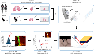
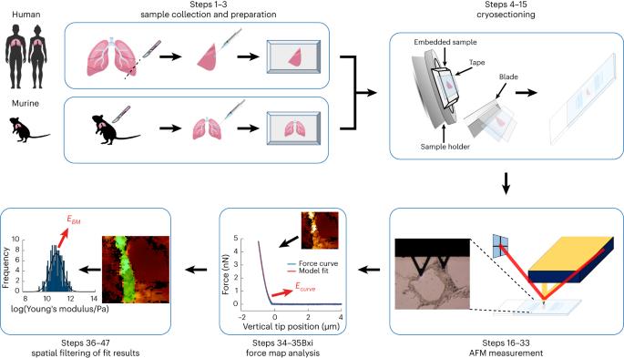
Mammalian cells sense and react to the mechanics of their immediate microenvironment. Therefore, the characterization of the biomechanical properties of tissues with high spatial resolution provides valuable insights into a broad variety of developmental, homeostatic and pathological processes within living organisms. The biomechanical properties of the basement membrane (BM), an extracellular matrix (ECM) substructure measuring only ∼100–400 nm across, are, among other things, pivotal to tumor progression and metastasis formation. Although the precise assignment of the Young’s modulus E of such a thin ECM substructure especially in between two cell layers is still challenging, biomechanical data of the BM can provide information of eminent diagnostic potential. Here we present a detailed protocol to quantify the elastic modulus of the BM in murine and human lung tissue, which is one of the major organs prone to metastasis. This protocol describes a streamlined workflow to determine the Young’s modulus E of the BM between the endothelial and epithelial cell layers shaping the alveolar wall in lung tissues using atomic force microscopy (AFM). Our step-by-step protocol provides instructions for murine and human lung tissue extraction, inflation of these tissues with cryogenic cutting medium, freezing and cryosectioning of the tissue samples, and AFM force-map recording. In addition, it guides the reader through a semi-automatic data analysis procedure to identify the pulmonary BM and extract its Young’s modulus E using an in-house tailored user-friendly AFM data analysis software, the Center for Applied Tissue Engineering and Regenerative Medicine processing toolbox, which enables automatic loading of the recorded force maps, conversion of the force versus piezo-extension curves to force versus indentation curves, calculation of Young’s moduli and generation of Young’s modulus maps, where the pulmonary BM can be identified using a semi-automatic spatial filtering tool. The entire protocol takes 1–2 d. Atomic force microscopy can be used to determine the stiffness of materials. This protocol describes how to measure and quantify the Young’s modulus E of pulmonary mouse and human basement membranes with atomic force microscopy and the Center for Applied Tissue Engineering and Regenerative Medicine processing toolbox.
期刊介绍:
Nature Protocols focuses on publishing protocols used to address significant biological and biomedical science research questions, including methods grounded in physics and chemistry with practical applications to biological problems. The journal caters to a primary audience of research scientists and, as such, exclusively publishes protocols with research applications. Protocols primarily aimed at influencing patient management and treatment decisions are not featured.
The specific techniques covered encompass a wide range, including but not limited to: Biochemistry, Cell biology, Cell culture, Chemical modification, Computational biology, Developmental biology, Epigenomics, Genetic analysis, Genetic modification, Genomics, Imaging, Immunology, Isolation, purification, and separation, Lipidomics, Metabolomics, Microbiology, Model organisms, Nanotechnology, Neuroscience, Nucleic-acid-based molecular biology, Pharmacology, Plant biology, Protein analysis, Proteomics, Spectroscopy, Structural biology, Synthetic chemistry, Tissue culture, Toxicology, and Virology.


 分享
分享
 求助内容:
求助内容: 应助结果提醒方式:
 扫码关注我们
