{"title":"Well-Differentiated Liposarcoma That Increased in Size after Menopause: A Case Report and a Review of the Literature.","authors":"Tatsuji Hoshino, Yoshihiro Takada, Ayako Sugihara, Masato Kinugasa, Yoshiyuki Tsuji","doi":"10.1155/2024/7599714","DOIUrl":null,"url":null,"abstract":"<p><p>This study reports a case of uterine liposarcoma together with a literature review. At 52 years old, our patient was diagnosed with lipoleiomyoma by MRI. A mass (39 × 32 × 41 mm<sup>3</sup>) protruding from the anterior wall of the uterine body was observed. When the patient was 58, her previous doctor found that the tumor had grown, and she was referred to the gynecology department of our hospital. On MRI, the major diameter was 1.23-fold longer and the volume was 1.85-fold higher compared with the prior imaging findings. Diffusion-weighted images revealed no significant anomalous signals. Thus, malignant tumors were included in the differential diagnosis. The patient consented to total abdominal hysterectomy and bilateral salpingo-oophorectomy. The mass on the anterior wall remained completely in the myometrium. No implantation was found in the abdominal cavity, and ascites was not detected. No bleeding or necrosis was observed on the cut surface. Histopathologically, differences in the sizes of adipocytes and stromal cells were identified. There were irregularities in the nuclear findings. The immunohistochemical findings were as follows: CDK4 (+), desmin (+), S100p (-), and Ki - 67 = 1%. Therefore, a diagnosis of well-differentiated liposarcoma was rendered. The lesion was localized in the uterus, and it was completely removed during surgery. Well-differentiated liposarcoma of uterine primary has no possibility of recurrence following complete resection, and thus, the patient underwent follow-up without additional treatment. No metastasis or recurrence has been observed for 10 months after surgery.</p>","PeriodicalId":9610,"journal":{"name":"Case Reports in Obstetrics and Gynecology","volume":"2024 ","pages":"7599714"},"PeriodicalIF":0.8000,"publicationDate":"2024-02-24","publicationTypes":"Journal Article","fieldsOfStudy":null,"isOpenAccess":false,"openAccessPdf":"https://www.ncbi.nlm.nih.gov/pmc/articles/PMC10908573/pdf/","citationCount":"0","resultStr":null,"platform":"Semanticscholar","paperid":null,"PeriodicalName":"Case Reports in Obstetrics and Gynecology","FirstCategoryId":"1085","ListUrlMain":"https://doi.org/10.1155/2024/7599714","RegionNum":0,"RegionCategory":null,"ArticlePicture":[],"TitleCN":null,"AbstractTextCN":null,"PMCID":null,"EPubDate":"2024/1/1 0:00:00","PubModel":"eCollection","JCR":"Q4","JCRName":"OBSTETRICS & GYNECOLOGY","Score":null,"Total":0}
引用次数: 0
Abstract
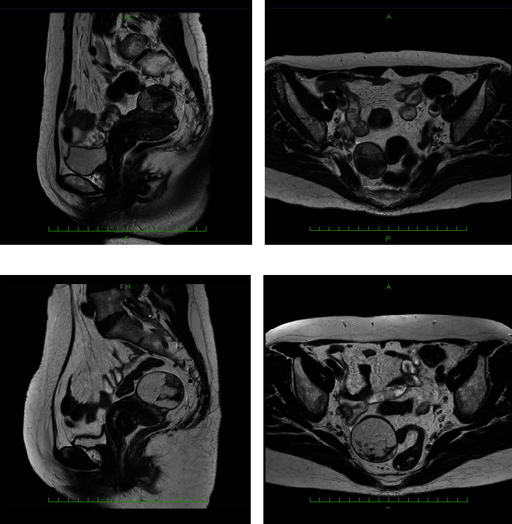
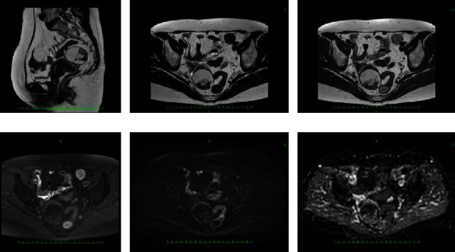
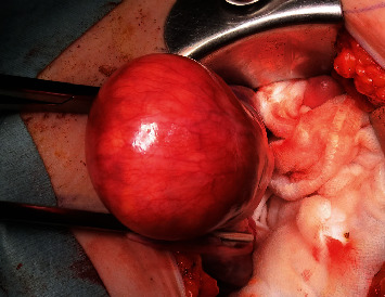
This study reports a case of uterine liposarcoma together with a literature review. At 52 years old, our patient was diagnosed with lipoleiomyoma by MRI. A mass (39 × 32 × 41 mm3) protruding from the anterior wall of the uterine body was observed. When the patient was 58, her previous doctor found that the tumor had grown, and she was referred to the gynecology department of our hospital. On MRI, the major diameter was 1.23-fold longer and the volume was 1.85-fold higher compared with the prior imaging findings. Diffusion-weighted images revealed no significant anomalous signals. Thus, malignant tumors were included in the differential diagnosis. The patient consented to total abdominal hysterectomy and bilateral salpingo-oophorectomy. The mass on the anterior wall remained completely in the myometrium. No implantation was found in the abdominal cavity, and ascites was not detected. No bleeding or necrosis was observed on the cut surface. Histopathologically, differences in the sizes of adipocytes and stromal cells were identified. There were irregularities in the nuclear findings. The immunohistochemical findings were as follows: CDK4 (+), desmin (+), S100p (-), and Ki - 67 = 1%. Therefore, a diagnosis of well-differentiated liposarcoma was rendered. The lesion was localized in the uterus, and it was completely removed during surgery. Well-differentiated liposarcoma of uterine primary has no possibility of recurrence following complete resection, and thus, the patient underwent follow-up without additional treatment. No metastasis or recurrence has been observed for 10 months after surgery.


 分享
分享
 求助内容:
求助内容: 应助结果提醒方式:
 扫码关注我们
