{"title":"CDC-like kinase 3 deficiency aggravates hypoxia-induced cardiomyocyte apoptosis through AKT signaling pathway.","authors":"Xiue Ma, Liming Gao, Rucun Ge, Tianyou Yuan, Bowen Lin, Lixiao Zhen","doi":"10.1007/s11626-024-00886-3","DOIUrl":null,"url":null,"abstract":"<p><p>Hypoxia-induced cardiomyocyte apoptosis is one major pathological change of acute myocardial infarction (AMI), but the underlying mechanism remains unexplored. CDC-like kinase 3 (CLK3) plays crucial roles in cell proliferation, migration and invasion, and nucleotide metabolism, however, the role of CLK3 in AMI, especially hypoxia-induced apoptosis, is largely unknown. The expression of CLK3 was elevated in mouse myocardial infarction (MI) models and neonatal rat ventricular myocytes (NRVMs) under hypoxia. Furthermore, CLK3 knockdown significantly promoted apoptosis and inhibited NRVM survival, while CLK3 overexpression promoted NRVM survival and inhibited apoptosis under hypoxic conditions. Mechanistically, CLK3 regulated the phosphorylation status of AKT, a key player in the regulation of apoptosis. Furthermore, overexpression of AKT rescued hypoxia-induced apoptosis in NRVMs caused by CLK3 deficiency. Taken together, CLK3 deficiency promotes hypoxia-induced cardiomyocyte apoptosis through AKT signaling pathway.</p>","PeriodicalId":13340,"journal":{"name":"In Vitro Cellular & Developmental Biology. Animal","volume":" ","pages":"333-342"},"PeriodicalIF":1.7000,"publicationDate":"2024-04-01","publicationTypes":"Journal Article","fieldsOfStudy":null,"isOpenAccess":false,"openAccessPdf":"","citationCount":"0","resultStr":null,"platform":"Semanticscholar","paperid":null,"PeriodicalName":"In Vitro Cellular & Developmental Biology. Animal","FirstCategoryId":"99","ListUrlMain":"https://doi.org/10.1007/s11626-024-00886-3","RegionNum":4,"RegionCategory":"生物学","ArticlePicture":[],"TitleCN":null,"AbstractTextCN":null,"PMCID":null,"EPubDate":"2024/3/4 0:00:00","PubModel":"Epub","JCR":"Q4","JCRName":"CELL BIOLOGY","Score":null,"Total":0}
引用次数: 0
Abstract
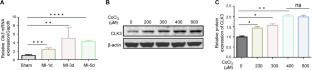
Hypoxia-induced cardiomyocyte apoptosis is one major pathological change of acute myocardial infarction (AMI), but the underlying mechanism remains unexplored. CDC-like kinase 3 (CLK3) plays crucial roles in cell proliferation, migration and invasion, and nucleotide metabolism, however, the role of CLK3 in AMI, especially hypoxia-induced apoptosis, is largely unknown. The expression of CLK3 was elevated in mouse myocardial infarction (MI) models and neonatal rat ventricular myocytes (NRVMs) under hypoxia. Furthermore, CLK3 knockdown significantly promoted apoptosis and inhibited NRVM survival, while CLK3 overexpression promoted NRVM survival and inhibited apoptosis under hypoxic conditions. Mechanistically, CLK3 regulated the phosphorylation status of AKT, a key player in the regulation of apoptosis. Furthermore, overexpression of AKT rescued hypoxia-induced apoptosis in NRVMs caused by CLK3 deficiency. Taken together, CLK3 deficiency promotes hypoxia-induced cardiomyocyte apoptosis through AKT signaling pathway.
期刊介绍:
In Vitro Cellular & Developmental Biology - Animal is a journal of the Society for In Vitro Biology (SIVB). Original manuscripts reporting results of research in cellular, molecular, and developmental biology that employ or are relevant to organs, tissue, tumors, and cells in vitro will be considered for publication. Topics covered include:
Biotechnology;
Cell and Tissue Models;
Cell Growth/Differentiation/Apoptosis;
Cellular Pathology/Virology;
Cytokines/Growth Factors/Adhesion Factors;
Establishment of Cell Lines;
Signal Transduction;
Stem Cells;
Toxicology/Chemical Carcinogenesis;
Product Applications.


 分享
分享
 求助内容:
求助内容: 应助结果提醒方式:
 扫码关注我们
