James A. Hendrixson, Alicen James, Nisreen S. Akel, Dominique J. Laster, Julie A. Crawford, Stuart B. Berryhill, Melda Onal
{"title":"Loss of chaperone-mediated autophagy does not alter age-related bone loss in male mice","authors":"James A. Hendrixson, Alicen James, Nisreen S. Akel, Dominique J. Laster, Julie A. Crawford, Stuart B. Berryhill, Melda Onal","doi":"10.1096/fba.2023-00133","DOIUrl":null,"url":null,"abstract":"<p>Chaperone-mediated autophagy (CMA) is a lysosome-dependent degradation pathway that eliminates proteins that are damaged, partially unfolded, or targeted for selective proteome remodeling. CMA contributes to several cellular processes, including stress response and proteostasis. Age-associated increase in cellular stressors and decrease in CMA contribute to pathologies associated with aging in various tissues. CMA contributes to bone homeostasis in young mice. An age-associated reduction in CMA was reported in osteoblast lineage cells; however, whether declining CMA contributes to skeletal aging is unknown. Herein we show that cellular stressors stimulate CMA in UAMS-32 osteoblastic cells. Moreover, the knockdown of an essential component of the CMA pathway, LAMP2A, sensitizes osteoblasts to cell death caused by DNA damage, ER stress, and oxidative stress. As elevations in these stressors are thought to contribute to age-related bone loss, we hypothesized that declining CMA contributes to the age-associated decline in bone formation by sensitizing osteoblast lineage cells to elevated stressors. To test this, we aged male CMA-deficient mice and controls up to 24 months of age and examined age-associated changes in bone mass and architecture. We showed that lack of CMA did not alter age-associated decline in bone mineral density as measured by dual x-ray absorptiometry (DXA). Moreover, microCT analysis performed at 24 months of age showed that vertebral cancellous bone volume, cortical thickness, and porosity of CMA-deficient and control mice were similar. Taken together, these results suggest that reduction of CMA does not contribute to age-related bone loss.</p>","PeriodicalId":12093,"journal":{"name":"FASEB bioAdvances","volume":"6 3","pages":"73-84"},"PeriodicalIF":2.0000,"publicationDate":"2024-02-20","publicationTypes":"Journal Article","fieldsOfStudy":null,"isOpenAccess":false,"openAccessPdf":"https://onlinelibrary.wiley.com/doi/epdf/10.1096/fba.2023-00133","citationCount":"0","resultStr":null,"platform":"Semanticscholar","paperid":null,"PeriodicalName":"FASEB bioAdvances","FirstCategoryId":"1085","ListUrlMain":"https://faseb.onlinelibrary.wiley.com/doi/10.1096/fba.2023-00133","RegionNum":0,"RegionCategory":null,"ArticlePicture":[],"TitleCN":null,"AbstractTextCN":null,"PMCID":null,"EPubDate":"","PubModel":"","JCR":"Q3","JCRName":"BIOCHEMISTRY & MOLECULAR BIOLOGY","Score":null,"Total":0}
引用次数: 0
Abstract
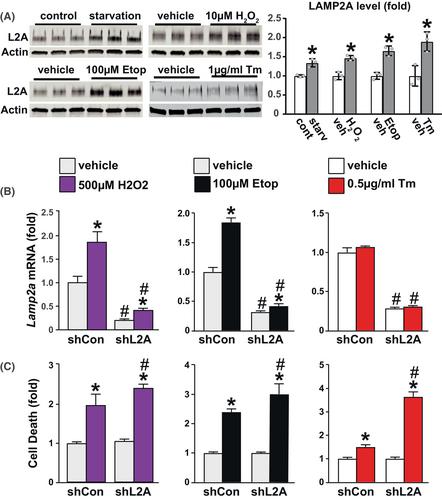
Chaperone-mediated autophagy (CMA) is a lysosome-dependent degradation pathway that eliminates proteins that are damaged, partially unfolded, or targeted for selective proteome remodeling. CMA contributes to several cellular processes, including stress response and proteostasis. Age-associated increase in cellular stressors and decrease in CMA contribute to pathologies associated with aging in various tissues. CMA contributes to bone homeostasis in young mice. An age-associated reduction in CMA was reported in osteoblast lineage cells; however, whether declining CMA contributes to skeletal aging is unknown. Herein we show that cellular stressors stimulate CMA in UAMS-32 osteoblastic cells. Moreover, the knockdown of an essential component of the CMA pathway, LAMP2A, sensitizes osteoblasts to cell death caused by DNA damage, ER stress, and oxidative stress. As elevations in these stressors are thought to contribute to age-related bone loss, we hypothesized that declining CMA contributes to the age-associated decline in bone formation by sensitizing osteoblast lineage cells to elevated stressors. To test this, we aged male CMA-deficient mice and controls up to 24 months of age and examined age-associated changes in bone mass and architecture. We showed that lack of CMA did not alter age-associated decline in bone mineral density as measured by dual x-ray absorptiometry (DXA). Moreover, microCT analysis performed at 24 months of age showed that vertebral cancellous bone volume, cortical thickness, and porosity of CMA-deficient and control mice were similar. Taken together, these results suggest that reduction of CMA does not contribute to age-related bone loss.


 分享
分享
 求助内容:
求助内容: 应助结果提醒方式:
 扫码关注我们
