{"title":"Correlation of laminin subunit alpha 3 expression in pancreatic ductal adenocarcinoma with tumor liver metastasis and survival.","authors":"Yueyi Xing, Xue Jing, Gong Qing, Yueping Jiang","doi":"10.2478/raon-2024-0020","DOIUrl":null,"url":null,"abstract":"<p><strong>Background: </strong>The high mortality rate of pancreatic ductal adenocarcinoma (PDAC) is primarily attributed to metastasis. Laminin subunit alpha 3 (LAMA3) is known to modulate tumor progression. However, the influence of LAMA3 on liver metastasis in PDAC remains unclear. This study aimed to elucidate whether LAMA3 expression is increased in PDAC with liver metastasis.</p><p><strong>Patients and methods: </strong>We extracted information related to LAMA3 expression levels and associated clinicopathological parameters from The Cancer Genome Atlas (TCGA) and four Gene Expression Omnibus (GEO) datasets. Clinicopathological analysis was performed; the Kaplan-Meier Plotter was used to evaluate LAMA3's prognostic effect in PDAC. We retrospectively collected clinicopathological data and tissue specimens from 117 surgically treated patients with PDAC at the Affiliated Hospital of Qingdao University. We assessed LAMA3 expression and investigated its correlation with the clinicopathological traits, clinical outcomes, and hepatic metastasis.</p><p><strong>Results: </strong>Amplified expression of LAMA3 was observed in PDAC tissue compared with normal tissue in the TCGA and GEO databases. High LAMA3 expression was associated with poor overall survival (OS) and relapse-free survival (RFS) in patients with PDAC. LAMA3 expression was significantly enhanced in PDAC tissues than in adjacent tissues. Tumor tissues from patients with PDAC exhibiting liver metastasis showed higher LAMA3 expression than those without liver metastasis. High LAMA3 expression correlated with large tumor size and TNM stage. LAMA3 expression and liver metastasis were independent predictive factors for OS; the former was independently associated with liver metastasis.</p><p><strong>Conclusions: </strong>LAMA3 expression is elevated in patients with PDAC with liver metastasis and is a predictor of prognosis.</p>","PeriodicalId":21034,"journal":{"name":"Radiology and Oncology","volume":" ","pages":"234-242"},"PeriodicalIF":2.2000,"publicationDate":"2024-03-07","publicationTypes":"Journal Article","fieldsOfStudy":null,"isOpenAccess":false,"openAccessPdf":"https://www.ncbi.nlm.nih.gov/pmc/articles/PMC11165973/pdf/","citationCount":"0","resultStr":null,"platform":"Semanticscholar","paperid":null,"PeriodicalName":"Radiology and Oncology","FirstCategoryId":"3","ListUrlMain":"https://doi.org/10.2478/raon-2024-0020","RegionNum":4,"RegionCategory":"医学","ArticlePicture":[],"TitleCN":null,"AbstractTextCN":null,"PMCID":null,"EPubDate":"2024/6/1 0:00:00","PubModel":"eCollection","JCR":"Q3","JCRName":"ONCOLOGY","Score":null,"Total":0}
引用次数: 0
Abstract
Background: The high mortality rate of pancreatic ductal adenocarcinoma (PDAC) is primarily attributed to metastasis. Laminin subunit alpha 3 (LAMA3) is known to modulate tumor progression. However, the influence of LAMA3 on liver metastasis in PDAC remains unclear. This study aimed to elucidate whether LAMA3 expression is increased in PDAC with liver metastasis.
Patients and methods: We extracted information related to LAMA3 expression levels and associated clinicopathological parameters from The Cancer Genome Atlas (TCGA) and four Gene Expression Omnibus (GEO) datasets. Clinicopathological analysis was performed; the Kaplan-Meier Plotter was used to evaluate LAMA3's prognostic effect in PDAC. We retrospectively collected clinicopathological data and tissue specimens from 117 surgically treated patients with PDAC at the Affiliated Hospital of Qingdao University. We assessed LAMA3 expression and investigated its correlation with the clinicopathological traits, clinical outcomes, and hepatic metastasis.
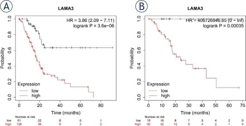
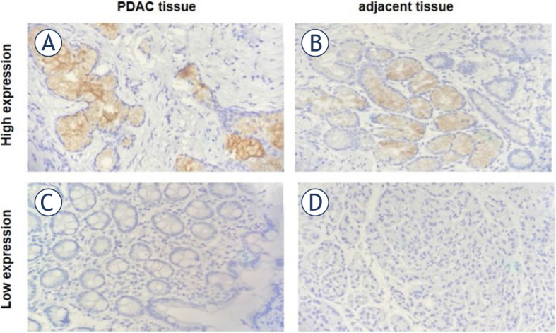
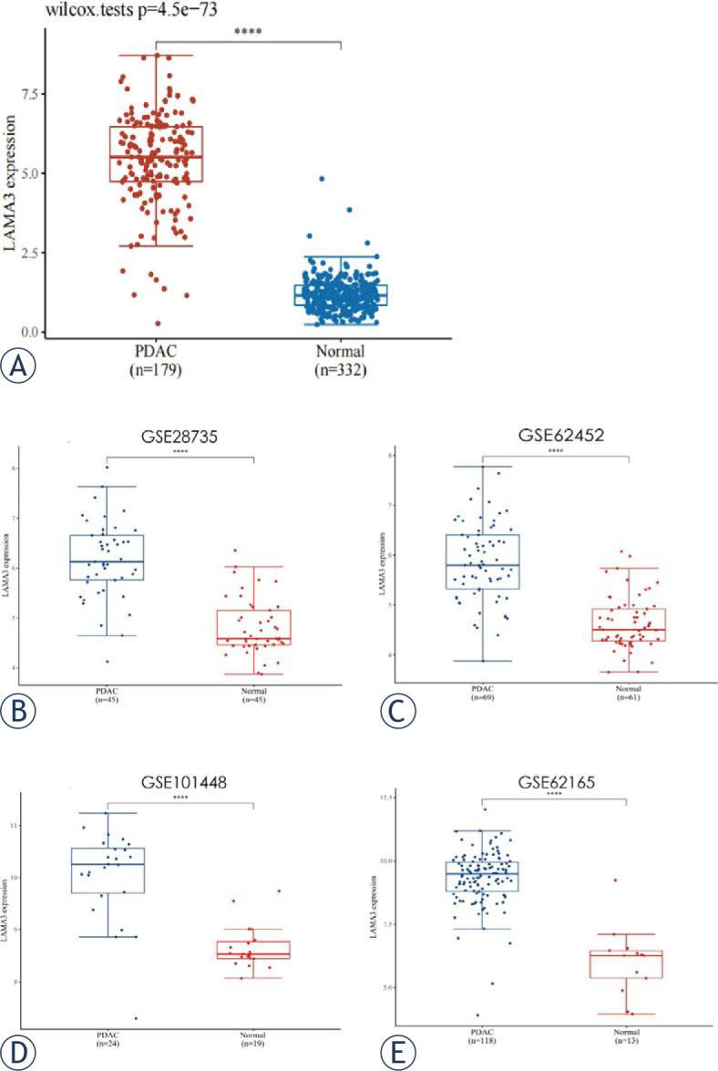
Results: Amplified expression of LAMA3 was observed in PDAC tissue compared with normal tissue in the TCGA and GEO databases. High LAMA3 expression was associated with poor overall survival (OS) and relapse-free survival (RFS) in patients with PDAC. LAMA3 expression was significantly enhanced in PDAC tissues than in adjacent tissues. Tumor tissues from patients with PDAC exhibiting liver metastasis showed higher LAMA3 expression than those without liver metastasis. High LAMA3 expression correlated with large tumor size and TNM stage. LAMA3 expression and liver metastasis were independent predictive factors for OS; the former was independently associated with liver metastasis.
Conclusions: LAMA3 expression is elevated in patients with PDAC with liver metastasis and is a predictor of prognosis.
期刊介绍:
Radiology and Oncology is a multidisciplinary journal devoted to the publishing original and high quality scientific papers and review articles, pertinent to diagnostic and interventional radiology, computerized tomography, magnetic resonance, ultrasound, nuclear medicine, radiotherapy, clinical and experimental oncology, radiobiology, medical physics and radiation protection. Therefore, the scope of the journal is to cover beside radiology the diagnostic and therapeutic aspects in oncology, which distinguishes it from other journals in the field.


 分享
分享
 求助内容:
求助内容: 应助结果提醒方式:
 扫码关注我们
