Franklin Guillermo Vargas-Corral, Américo Ernesto Vargas-Corral, Miguel Angel Rodríguez-Valverde, Manuel Bravo, Juan Ignacio Rosales-Leal
{"title":"Clinical comparison of marginal fit of ceramic inlays between digital and conventional impressions.","authors":"Franklin Guillermo Vargas-Corral, Américo Ernesto Vargas-Corral, Miguel Angel Rodríguez-Valverde, Manuel Bravo, Juan Ignacio Rosales-Leal","doi":"10.4047/jap.2024.16.1.57","DOIUrl":null,"url":null,"abstract":"<p><strong>Purpose: </strong>The aim of this stuldy was to compare the clinical marginal fit of CAD-CAM inlays obtained from intraoral digital impression or addition silicone impression techniques.</p><p><strong>Materials and methods: </strong>The study included 31 inlays for prosthodontics purposes of 31 patients: 15 based on intraoral digital impressions (DI group); and 16 based on a conventional impression technique (CI group). Inlays included occlusal and a non-occlusal surface. Inlays were milled in ceramic. The inlay-teeth interface was replicated by placing each inlay in its corresponding uncemented clinical preparation and taking interface impressions with silicone material from occlusal and free surfaces. Interface analysis was made using white light confocal microscopy (WLCM) (scanning area: 694 × 510 µm<sup>2</sup>) from the impression samples. The gap size and the inlay overextension were measured from the microscopy topographies. For analytical purposes (i.e., 95-%-confidence intervals calculations and <i>P</i>-value calculations), the procedure REGRESS in SUDAAN was used to account for clustering (i.e., multiple measurements). For p-value calculation, the log transformation of the dependent variables was used to normalize the distributions.</p><p><strong>Results: </strong>Marginal fit values for occlusal and free surfaces were affected by the type of impression. There were no differences between surfaces (occlusal vs. free). Gap obtained for DI group was 164 ± 84 µm and that for CI group was 209 ± 104 µm, and there were statistical differences between them (<i>p</i> = .041). Mean overextension values were 60 ± 59 µm for DI group and 67 ± 73 µm for CI group, and there were no differences between then (<i>p</i> = .553).</p><p><strong>Conclusion: </strong>Digital impression achieved inlays with higher clinical marginal fit and performed better than the conventional silicone materials.</p>","PeriodicalId":51291,"journal":{"name":"Journal of Advanced Prosthodontics","volume":"16 1","pages":"57-65"},"PeriodicalIF":2.5000,"publicationDate":"2024-02-01","publicationTypes":"Journal Article","fieldsOfStudy":null,"isOpenAccess":false,"openAccessPdf":"https://www.ncbi.nlm.nih.gov/pmc/articles/PMC10917630/pdf/","citationCount":"0","resultStr":null,"platform":"Semanticscholar","paperid":null,"PeriodicalName":"Journal of Advanced Prosthodontics","FirstCategoryId":"3","ListUrlMain":"https://doi.org/10.4047/jap.2024.16.1.57","RegionNum":3,"RegionCategory":"医学","ArticlePicture":[],"TitleCN":null,"AbstractTextCN":null,"PMCID":null,"EPubDate":"2024/2/23 0:00:00","PubModel":"Epub","JCR":"Q1","JCRName":"DENTISTRY, ORAL SURGERY & MEDICINE","Score":null,"Total":0}
引用次数: 0
Abstract
Purpose: The aim of this stuldy was to compare the clinical marginal fit of CAD-CAM inlays obtained from intraoral digital impression or addition silicone impression techniques.
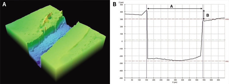
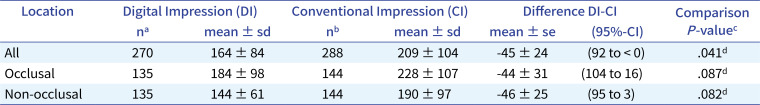
Materials and methods: The study included 31 inlays for prosthodontics purposes of 31 patients: 15 based on intraoral digital impressions (DI group); and 16 based on a conventional impression technique (CI group). Inlays included occlusal and a non-occlusal surface. Inlays were milled in ceramic. The inlay-teeth interface was replicated by placing each inlay in its corresponding uncemented clinical preparation and taking interface impressions with silicone material from occlusal and free surfaces. Interface analysis was made using white light confocal microscopy (WLCM) (scanning area: 694 × 510 µm2) from the impression samples. The gap size and the inlay overextension were measured from the microscopy topographies. For analytical purposes (i.e., 95-%-confidence intervals calculations and P-value calculations), the procedure REGRESS in SUDAAN was used to account for clustering (i.e., multiple measurements). For p-value calculation, the log transformation of the dependent variables was used to normalize the distributions.
Results: Marginal fit values for occlusal and free surfaces were affected by the type of impression. There were no differences between surfaces (occlusal vs. free). Gap obtained for DI group was 164 ± 84 µm and that for CI group was 209 ± 104 µm, and there were statistical differences between them (p = .041). Mean overextension values were 60 ± 59 µm for DI group and 67 ± 73 µm for CI group, and there were no differences between then (p = .553).
Conclusion: Digital impression achieved inlays with higher clinical marginal fit and performed better than the conventional silicone materials.
期刊介绍:
This journal aims to convey scientific and clinical progress in the field of prosthodontics and its related areas to many dental communities concerned with esthetic and functional restorations, occlusion, implants, prostheses, and biomaterials related to prosthodontics.
This journal publishes
• Original research data of high scientific merit in the field of diagnosis, function, esthetics and stomatognathic physiology related to prosthodontic rehabilitation, physiology and mechanics of occlusion, mechanical and biologic aspects of prosthodontic materials including dental implants.
• Review articles by experts on controversies and new developments in prosthodontics.
• Case reports if they provide or document new fundamental knowledge.


 分享
分享
 求助内容:
求助内容: 应助结果提醒方式:
 扫码关注我们
