Liang Jiang, Jiarui Sun, Yajing Wang, Haodi Yang, Yu-Chen Chen, Mingyang Peng, Hong Zhang, Yang Chen, Xindao Yin
{"title":"Diffusion-/perfusion-weighted imaging fusion to automatically identify stroke within 4.5 h.","authors":"Liang Jiang, Jiarui Sun, Yajing Wang, Haodi Yang, Yu-Chen Chen, Mingyang Peng, Hong Zhang, Yang Chen, Xindao Yin","doi":"10.1007/s00330-024-10619-5","DOIUrl":null,"url":null,"abstract":"<p><strong>Objectives: </strong>We aimed to develop machine learning (ML) models based on diffusion- and perfusion-weighted imaging fusion (DP fusion) for identifying stroke within 4.5 h, to compare them with DWI- and/or PWI-based ML models, and to construct an automatic segmentation-classification model and compare with manual labeling methods.</p><p><strong>Methods: </strong>ML models were developed from multimodal MRI datasets of acute stroke patients within 24 h of clear symptom onset from two centers. The processes included manual segmentation, registration, DP fusion, feature extraction, and model establishment (logistic regression (LR) and support vector machine (SVM)). A segmentation-classification model (X-Net) was proposed for automatically identifying stroke within 4.5 h. The area under the receiver operating characteristic curve (AUC), sensitivity, Dice coefficients, decision curve analysis, and calibration curves were used to evaluate model performance.</p><p><strong>Results: </strong>A total of 418 patients (≤ 4.5 h: 214; > 4.5 h: 204) were evaluated. The DP fusion model achieved the highest AUC in identifying the onset time in the training (LR: 0.95; SVM: 0.92) and test sets (LR: 0.91; SVM: 0.90). The DP fusion-LR model displayed consistent positive and greater net benefits than other models across a broad range of risk thresholds. The calibration curve demonstrated the good calibration of the DP fusion-LR model (average absolute error: 0.049). The X-Net model obtained the highest Dice coefficients (DWI: 0.81; Tmax: 0.83) and achieved similar performance to manual labeling (AUC: 0.84).</p><p><strong>Conclusions: </strong>The automatic segmentation-classification models based on DWI and PWI fusion images had high performance in identifying stroke within 4.5 h.</p><p><strong>Clinical relevance statement: </strong>Perfusion-weighted imaging (PWI) fusion images had high performance in identifying stroke within 4.5 h. The automatic segmentation-classification models based on DWI and PWI fusion images could provide clinicians with decision-making guidance for acute stroke patients with unknown onset time.</p><p><strong>Key points: </strong>• The diffusion/perfusion-weighted imaging fusion model had the best performance in identifying stroke within 4.5 h. • The X-Net model had the highest Dice and achieved performance close to manual labeling in segmenting lesions of acute stroke. • The automatic segmentation-classification model based on DP fusion images performed well in identifying stroke within 4.5 h.</p>","PeriodicalId":12076,"journal":{"name":"European Radiology","volume":" ","pages":"6808-6819"},"PeriodicalIF":4.7000,"publicationDate":"2024-10-01","publicationTypes":"Journal Article","fieldsOfStudy":null,"isOpenAccess":false,"openAccessPdf":"https://www.ncbi.nlm.nih.gov/pmc/articles/PMC11399198/pdf/","citationCount":"0","resultStr":null,"platform":"Semanticscholar","paperid":null,"PeriodicalName":"European Radiology","FirstCategoryId":"3","ListUrlMain":"https://doi.org/10.1007/s00330-024-10619-5","RegionNum":2,"RegionCategory":"医学","ArticlePicture":[],"TitleCN":null,"AbstractTextCN":null,"PMCID":null,"EPubDate":"2024/3/15 0:00:00","PubModel":"Epub","JCR":"Q1","JCRName":"RADIOLOGY, NUCLEAR MEDICINE & MEDICAL IMAGING","Score":null,"Total":0}
引用次数: 0
Abstract
Objectives: We aimed to develop machine learning (ML) models based on diffusion- and perfusion-weighted imaging fusion (DP fusion) for identifying stroke within 4.5 h, to compare them with DWI- and/or PWI-based ML models, and to construct an automatic segmentation-classification model and compare with manual labeling methods.
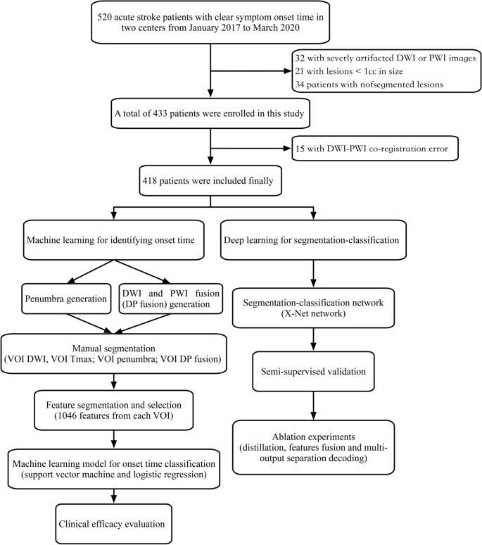
Methods: ML models were developed from multimodal MRI datasets of acute stroke patients within 24 h of clear symptom onset from two centers. The processes included manual segmentation, registration, DP fusion, feature extraction, and model establishment (logistic regression (LR) and support vector machine (SVM)). A segmentation-classification model (X-Net) was proposed for automatically identifying stroke within 4.5 h. The area under the receiver operating characteristic curve (AUC), sensitivity, Dice coefficients, decision curve analysis, and calibration curves were used to evaluate model performance.
Results: A total of 418 patients (≤ 4.5 h: 214; > 4.5 h: 204) were evaluated. The DP fusion model achieved the highest AUC in identifying the onset time in the training (LR: 0.95; SVM: 0.92) and test sets (LR: 0.91; SVM: 0.90). The DP fusion-LR model displayed consistent positive and greater net benefits than other models across a broad range of risk thresholds. The calibration curve demonstrated the good calibration of the DP fusion-LR model (average absolute error: 0.049). The X-Net model obtained the highest Dice coefficients (DWI: 0.81; Tmax: 0.83) and achieved similar performance to manual labeling (AUC: 0.84).
Conclusions: The automatic segmentation-classification models based on DWI and PWI fusion images had high performance in identifying stroke within 4.5 h.
Clinical relevance statement: Perfusion-weighted imaging (PWI) fusion images had high performance in identifying stroke within 4.5 h. The automatic segmentation-classification models based on DWI and PWI fusion images could provide clinicians with decision-making guidance for acute stroke patients with unknown onset time.
Key points: • The diffusion/perfusion-weighted imaging fusion model had the best performance in identifying stroke within 4.5 h. • The X-Net model had the highest Dice and achieved performance close to manual labeling in segmenting lesions of acute stroke. • The automatic segmentation-classification model based on DP fusion images performed well in identifying stroke within 4.5 h.
期刊介绍:
European Radiology (ER) continuously updates scientific knowledge in radiology by publication of strong original articles and state-of-the-art reviews written by leading radiologists. A well balanced combination of review articles, original papers, short communications from European radiological congresses and information on society matters makes ER an indispensable source for current information in this field.
This is the Journal of the European Society of Radiology, and the official journal of a number of societies.
From 2004-2008 supplements to European Radiology were published under its companion, European Radiology Supplements, ISSN 1613-3749.


 分享
分享
 求助内容:
求助内容: 应助结果提醒方式:
 扫码关注我们
