{"title":"Synthetic MRI and amide proton transfer-weighted MRI for differentiating between benign and malignant sinonasal lesions.","authors":"Ying Xiang, Qiujuan Zhang, Xin Chen, Honghong Sun, Xiaohui Li, Xiaocheng Wei, Jinman Zhong, Bo Gao, Wei Huang, Wenbin Liang, Haiqiao Sun, Quanxin Yang, Xiaoyong Ren","doi":"10.1007/s00330-024-10696-6","DOIUrl":null,"url":null,"abstract":"<p><strong>Objectives: </strong>To explore the value of the synthetic MRI (SyMRI), combined with amide proton transfer-weighted (APTw) MRI for quantitative and morphologic assessment of sinonasal lesions, which could provide relative scale for the quantitative assessment of tissue properties.</p><p><strong>Methods: </strong>A total of 80 patients (31 malignant and 49 benign) with sinonasal lesions, who underwent the SyMRI and APTw examination, were retrospectively analyzed. Quantitative parameters (T1, T2, proton density (PD)) and APT % were obtained through outlining the region of interest (ROI) and comparing the two groups utilizing independent Student t test or a Wilcoxon test. Receiver operating characteristic curve (ROC), Delong test, and logistic regression analysis were performed to assess the diagnostic efficiency of one-parameter and multiparametric models.</p><p><strong>Results: </strong>SyMRI-derived mean T1, T2, and PD were significantly higher and APT % was relatively lower in benign compared to malignant sinonasal lesions (p < 0.05). The ROC analysis showed that the AUCs of the SyMRI-derived quantitative (T1, T2, PD) values and APT % ranged from 0.677 to 0.781 for differential diagnosis between benign and malignant sinonasal lesions. The T2 values showed the best diagnostic performance among all single parameters for differentiating these two masses. The AUCs of combined SyMRI-derived multiple parameters with APT % (AUC = 0.866) were the highest than that of any single parameter, which was significantly improved (p < 0.05).</p><p><strong>Conclusion: </strong>The combination of SyMRI and APTw imaging has the potential to reflect intrinsic tissue characteristics useful for differentiating benign from malignant sinonasal lesions.</p><p><strong>Clinical relevance statement: </strong>Combining synthetic MRI with amide proton transfer-weighted imaging could function as a quantitative and contrast-free approach, significantly enhancing the differentiation of benign and malignant sinonasal lesions and overcoming the limitations associated with the superficial nature of endoscopic nasal sampling.</p><p><strong>Key points: </strong>• Synthetic MRI and amide proton transfer-weighted MRI could differentiate benign from malignant sinonasal lesions based on quantitative parameters. • The diagnostic efficiency could be significantly improved through synthetic MRI + amide proton transfer-weighted imaging. • The combination of synthetic MRI and amide proton transfer-weighted MRI is a noninvasive method to evaluate sinonasal lesions.</p>","PeriodicalId":12076,"journal":{"name":"European Radiology","volume":" ","pages":"6820-6830"},"PeriodicalIF":4.7000,"publicationDate":"2024-10-01","publicationTypes":"Journal Article","fieldsOfStudy":null,"isOpenAccess":false,"openAccessPdf":"","citationCount":"0","resultStr":null,"platform":"Semanticscholar","paperid":null,"PeriodicalName":"European Radiology","FirstCategoryId":"3","ListUrlMain":"https://doi.org/10.1007/s00330-024-10696-6","RegionNum":2,"RegionCategory":"医学","ArticlePicture":[],"TitleCN":null,"AbstractTextCN":null,"PMCID":null,"EPubDate":"2024/3/16 0:00:00","PubModel":"Epub","JCR":"Q1","JCRName":"RADIOLOGY, NUCLEAR MEDICINE & MEDICAL IMAGING","Score":null,"Total":0}
引用次数: 0
Abstract
Objectives: To explore the value of the synthetic MRI (SyMRI), combined with amide proton transfer-weighted (APTw) MRI for quantitative and morphologic assessment of sinonasal lesions, which could provide relative scale for the quantitative assessment of tissue properties.
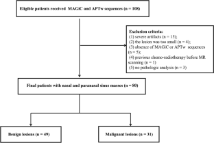
Methods: A total of 80 patients (31 malignant and 49 benign) with sinonasal lesions, who underwent the SyMRI and APTw examination, were retrospectively analyzed. Quantitative parameters (T1, T2, proton density (PD)) and APT % were obtained through outlining the region of interest (ROI) and comparing the two groups utilizing independent Student t test or a Wilcoxon test. Receiver operating characteristic curve (ROC), Delong test, and logistic regression analysis were performed to assess the diagnostic efficiency of one-parameter and multiparametric models.
Results: SyMRI-derived mean T1, T2, and PD were significantly higher and APT % was relatively lower in benign compared to malignant sinonasal lesions (p < 0.05). The ROC analysis showed that the AUCs of the SyMRI-derived quantitative (T1, T2, PD) values and APT % ranged from 0.677 to 0.781 for differential diagnosis between benign and malignant sinonasal lesions. The T2 values showed the best diagnostic performance among all single parameters for differentiating these two masses. The AUCs of combined SyMRI-derived multiple parameters with APT % (AUC = 0.866) were the highest than that of any single parameter, which was significantly improved (p < 0.05).
Conclusion: The combination of SyMRI and APTw imaging has the potential to reflect intrinsic tissue characteristics useful for differentiating benign from malignant sinonasal lesions.
Clinical relevance statement: Combining synthetic MRI with amide proton transfer-weighted imaging could function as a quantitative and contrast-free approach, significantly enhancing the differentiation of benign and malignant sinonasal lesions and overcoming the limitations associated with the superficial nature of endoscopic nasal sampling.
Key points: • Synthetic MRI and amide proton transfer-weighted MRI could differentiate benign from malignant sinonasal lesions based on quantitative parameters. • The diagnostic efficiency could be significantly improved through synthetic MRI + amide proton transfer-weighted imaging. • The combination of synthetic MRI and amide proton transfer-weighted MRI is a noninvasive method to evaluate sinonasal lesions.
期刊介绍:
European Radiology (ER) continuously updates scientific knowledge in radiology by publication of strong original articles and state-of-the-art reviews written by leading radiologists. A well balanced combination of review articles, original papers, short communications from European radiological congresses and information on society matters makes ER an indispensable source for current information in this field.
This is the Journal of the European Society of Radiology, and the official journal of a number of societies.
From 2004-2008 supplements to European Radiology were published under its companion, European Radiology Supplements, ISSN 1613-3749.


 分享
分享
 求助内容:
求助内容: 应助结果提醒方式:
 扫码关注我们
