Valencia Hui Xian Foo, Gilbert Y S Lim, Yu-Chi Liu, Hon Shing Ong, Evan Wong, Stacy Chan, Jipson Wong, Jodhbir S Mehta, Daniel S W Ting, Marcus Ang
{"title":"Deep learning for detection of Fuchs endothelial dystrophy from widefield specular microscopy imaging: a pilot study.","authors":"Valencia Hui Xian Foo, Gilbert Y S Lim, Yu-Chi Liu, Hon Shing Ong, Evan Wong, Stacy Chan, Jipson Wong, Jodhbir S Mehta, Daniel S W Ting, Marcus Ang","doi":"10.1186/s40662-024-00378-1","DOIUrl":null,"url":null,"abstract":"<p><strong>Background: </strong>To describe the diagnostic performance of a deep learning (DL) algorithm in detecting Fuchs endothelial corneal dystrophy (FECD) based on specular microscopy (SM) and to reliably detect widefield peripheral SM images with an endothelial cell density (ECD) > 1000 cells/mm<sup>2</sup>.</p><p><strong>Methods: </strong>Five hundred and forty-seven subjects had SM imaging performed for the central cornea endothelium. One hundred and seventy-three images had FECD, while 602 images had other diagnoses. Using fivefold cross-validation on the dataset containing 775 central SM images combined with ECD, coefficient of variation (CV) and hexagonal endothelial cell ratio (HEX), the first DL model was trained to discriminate FECD from other images and was further tested on an external set of 180 images. In eyes with FECD, a separate DL model was trained with 753 central/paracentral SM images to detect SM with ECD > 1000 cells/mm<sup>2</sup> and tested on 557 peripheral SM images. Area under curve (AUC), sensitivity and specificity were evaluated.</p><p><strong>Results: </strong>The first model achieved an AUC of 0.96 with 0.91 sensitivity and 0.91 specificity in detecting FECD from other images. With an external validation set, the model achieved an AUC of 0.77, with a sensitivity of 0.69 and specificity of 0.68 in differentiating FECD from other diagnoses. The second model achieved an AUC of 0.88 with 0.79 sensitivity and 0.78 specificity in detecting peripheral SM images with ECD > 1000 cells/mm<sup>2</sup>.</p><p><strong>Conclusions: </strong>Our pilot study developed a DL model that could reliably detect FECD from other SM images and identify widefield SM images with ECD > 1000 cells/mm<sup>2</sup> in eyes with FECD. This could be the foundation for future DL models to track progression of eyes with FECD and identify candidates suitable for therapies such as Descemet stripping only.</p>","PeriodicalId":12194,"journal":{"name":"Eye and Vision","volume":"11 1","pages":"11"},"PeriodicalIF":4.0000,"publicationDate":"2024-03-18","publicationTypes":"Journal Article","fieldsOfStudy":null,"isOpenAccess":false,"openAccessPdf":"https://www.ncbi.nlm.nih.gov/pmc/articles/PMC10946096/pdf/","citationCount":"0","resultStr":null,"platform":"Semanticscholar","paperid":null,"PeriodicalName":"Eye and Vision","FirstCategoryId":"3","ListUrlMain":"https://doi.org/10.1186/s40662-024-00378-1","RegionNum":1,"RegionCategory":"医学","ArticlePicture":[],"TitleCN":null,"AbstractTextCN":null,"PMCID":null,"EPubDate":"","PubModel":"","JCR":"Q1","JCRName":"OPHTHALMOLOGY","Score":null,"Total":0}
引用次数: 0
Abstract
Background: To describe the diagnostic performance of a deep learning (DL) algorithm in detecting Fuchs endothelial corneal dystrophy (FECD) based on specular microscopy (SM) and to reliably detect widefield peripheral SM images with an endothelial cell density (ECD) > 1000 cells/mm2.
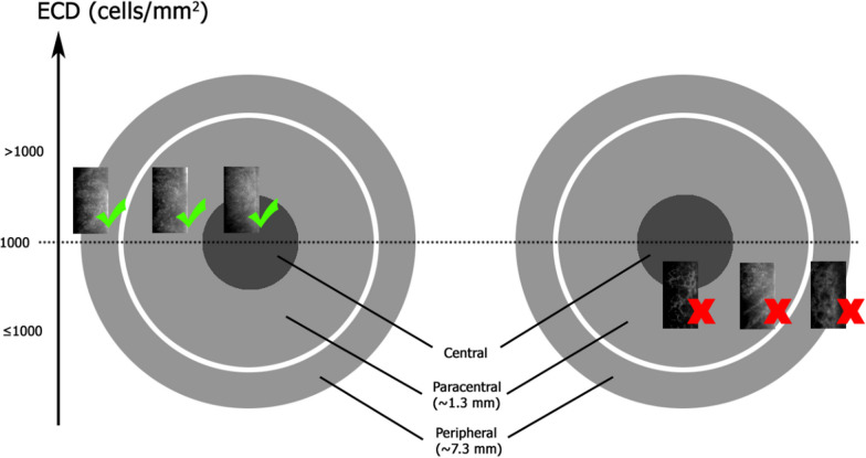
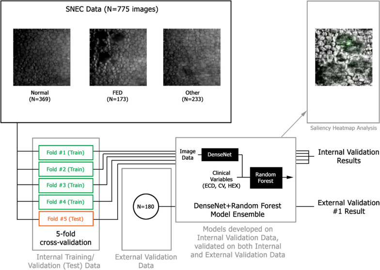
Methods: Five hundred and forty-seven subjects had SM imaging performed for the central cornea endothelium. One hundred and seventy-three images had FECD, while 602 images had other diagnoses. Using fivefold cross-validation on the dataset containing 775 central SM images combined with ECD, coefficient of variation (CV) and hexagonal endothelial cell ratio (HEX), the first DL model was trained to discriminate FECD from other images and was further tested on an external set of 180 images. In eyes with FECD, a separate DL model was trained with 753 central/paracentral SM images to detect SM with ECD > 1000 cells/mm2 and tested on 557 peripheral SM images. Area under curve (AUC), sensitivity and specificity were evaluated.
Results: The first model achieved an AUC of 0.96 with 0.91 sensitivity and 0.91 specificity in detecting FECD from other images. With an external validation set, the model achieved an AUC of 0.77, with a sensitivity of 0.69 and specificity of 0.68 in differentiating FECD from other diagnoses. The second model achieved an AUC of 0.88 with 0.79 sensitivity and 0.78 specificity in detecting peripheral SM images with ECD > 1000 cells/mm2.
Conclusions: Our pilot study developed a DL model that could reliably detect FECD from other SM images and identify widefield SM images with ECD > 1000 cells/mm2 in eyes with FECD. This could be the foundation for future DL models to track progression of eyes with FECD and identify candidates suitable for therapies such as Descemet stripping only.
期刊介绍:
Eye and Vision is an open access, peer-reviewed journal for ophthalmologists and visual science specialists. It welcomes research articles, reviews, methodologies, commentaries, case reports, perspectives and short reports encompassing all aspects of eye and vision. Topics of interest include but are not limited to: current developments of theoretical, experimental and clinical investigations in ophthalmology, optometry and vision science which focus on novel and high-impact findings on central issues pertaining to biology, pathophysiology and etiology of eye diseases as well as advances in diagnostic techniques, surgical treatment, instrument updates, the latest drug findings, results of clinical trials and research findings. It aims to provide ophthalmologists and visual science specialists with the latest developments in theoretical, experimental and clinical investigations in eye and vision.


 分享
分享
 求助内容:
求助内容: 应助结果提醒方式:
 扫码关注我们
