Doppler ultrasound-based evaluation of hemodynamic changes in the ophthalmic artery and central retinal artery in patients with type 1 diabetes mellitus without retinopathy and with mild non-proliferative retinopathy.
{"title":"Doppler ultrasound-based evaluation of hemodynamic changes in the ophthalmic artery and central retinal artery in patients with type 1 diabetes mellitus without retinopathy and with mild non-proliferative retinopathy.","authors":"Magdalena Pauk-Domańska, Agnieszka Wilczewska, Dominika Jaguś, Bartosz Kaczyński, Wiesław Jakubowski","doi":"10.15557/jou.2024.0009","DOIUrl":null,"url":null,"abstract":"<p><strong>Aim: </strong>Determination of blood flow parameters in the ophthalmic artery and central retinal artery using Doppler ultrasound in patients with type 1 diabetes mellitus without fundus signs of diabetic retinopathy and with mild non-proliferative retinopathy.</p><p><strong>Material and methods: </strong>To eliminate the impact of other systemic factors on vascular flow, the study enrolled a total of 80 patients with type 1 diabetes mellitus, aged between 18 and 45 years. The study participants did not have any diabetic complications or other systemic or ocular comorbidities. The control group comprised 81 healthy individuals within a similar age range. Color Doppler ultrasound examinations of the ophthalmic artery and central retinal artery were performed to evaluate selected blood flow parameters including peak systolic velocity, end-diastolic velocity, and resistance index.</p><p><strong>Results: </strong>Patients with type 1 diabetes mellitus exhibited statistically significant decrease in both systolic and end-diastolic velocities in the central retinal artery, accompanied by an elevation in resistance index, compared to the control group. The study revealed differences in blood flow parameters between the patients without fundus changes and those exhibiting mild non-proliferative retinopathy. Specifically, patients with retinopathy showed a significant decrease in both systolic velocity and end-diastolic velocity in the central retinal artery. No differences were observed for the same parameters in the ophthalmic artery. When analyzing the patients' blood flow parameters in relation to the degree of diabetes control, as determined by glycated hemoglobin levels, a statistically significant reduction in systolic velocity was identified in both the ophthalmic and central retinal arteries in the group with poorly controlled diabetes.</p><p><strong>Conclusions: </strong>Examination of the orbital vessels using Doppler ultrasound in patients with type 1 diabetes mellitus holds promise as an effective method for early detection of vascular abnormalities.</p>","PeriodicalId":45612,"journal":{"name":"Journal of Ultrasonography","volume":"24 96","pages":"20240009"},"PeriodicalIF":1.5000,"publicationDate":"2024-03-14","publicationTypes":"Journal Article","fieldsOfStudy":null,"isOpenAccess":false,"openAccessPdf":"https://www.ncbi.nlm.nih.gov/pmc/articles/PMC10940271/pdf/","citationCount":"0","resultStr":null,"platform":"Semanticscholar","paperid":null,"PeriodicalName":"Journal of Ultrasonography","FirstCategoryId":"1085","ListUrlMain":"https://doi.org/10.15557/jou.2024.0009","RegionNum":0,"RegionCategory":null,"ArticlePicture":[],"TitleCN":null,"AbstractTextCN":null,"PMCID":null,"EPubDate":"2024/2/1 0:00:00","PubModel":"eCollection","JCR":"Q3","JCRName":"RADIOLOGY, NUCLEAR MEDICINE & MEDICAL IMAGING","Score":null,"Total":0}
引用次数: 0
Abstract
Aim: Determination of blood flow parameters in the ophthalmic artery and central retinal artery using Doppler ultrasound in patients with type 1 diabetes mellitus without fundus signs of diabetic retinopathy and with mild non-proliferative retinopathy.
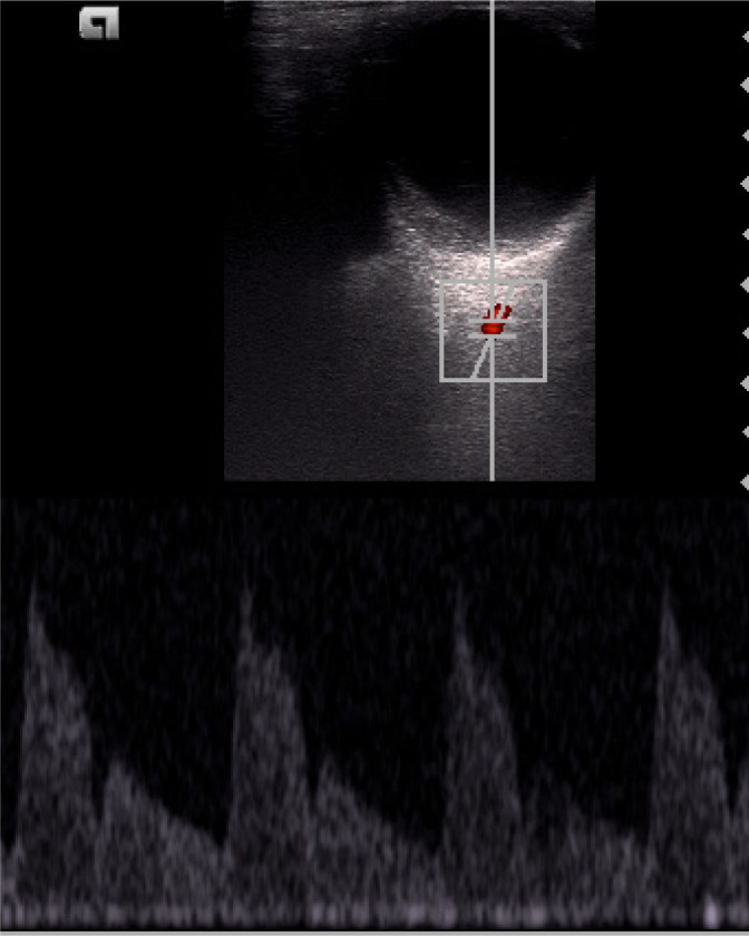
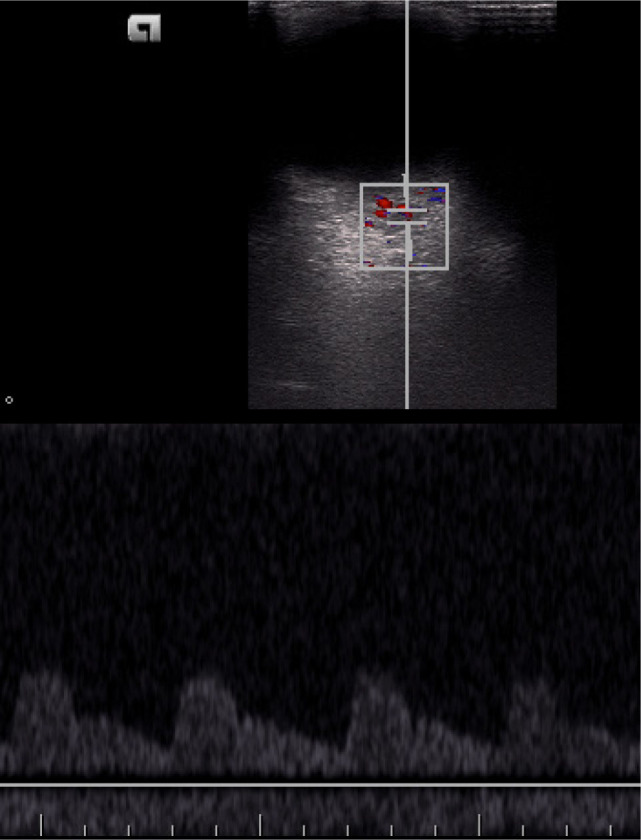
Material and methods: To eliminate the impact of other systemic factors on vascular flow, the study enrolled a total of 80 patients with type 1 diabetes mellitus, aged between 18 and 45 years. The study participants did not have any diabetic complications or other systemic or ocular comorbidities. The control group comprised 81 healthy individuals within a similar age range. Color Doppler ultrasound examinations of the ophthalmic artery and central retinal artery were performed to evaluate selected blood flow parameters including peak systolic velocity, end-diastolic velocity, and resistance index.
Results: Patients with type 1 diabetes mellitus exhibited statistically significant decrease in both systolic and end-diastolic velocities in the central retinal artery, accompanied by an elevation in resistance index, compared to the control group. The study revealed differences in blood flow parameters between the patients without fundus changes and those exhibiting mild non-proliferative retinopathy. Specifically, patients with retinopathy showed a significant decrease in both systolic velocity and end-diastolic velocity in the central retinal artery. No differences were observed for the same parameters in the ophthalmic artery. When analyzing the patients' blood flow parameters in relation to the degree of diabetes control, as determined by glycated hemoglobin levels, a statistically significant reduction in systolic velocity was identified in both the ophthalmic and central retinal arteries in the group with poorly controlled diabetes.
Conclusions: Examination of the orbital vessels using Doppler ultrasound in patients with type 1 diabetes mellitus holds promise as an effective method for early detection of vascular abnormalities.


 分享
分享
 求助内容:
求助内容: 应助结果提醒方式:
 扫码关注我们
