Isil Merve Torun, Taha Baysal, Mirac Aysen Unsal, Murat Sonmez
{"title":"Choroid and Retinal Effects of Epilepsy and Epilepsy Subgroups.","authors":"Isil Merve Torun, Taha Baysal, Mirac Aysen Unsal, Murat Sonmez","doi":"10.14744/bej.2023.19942","DOIUrl":null,"url":null,"abstract":"<p><strong>Objectives: </strong>The objective were to evaluate structural alterations in the retina and choroid tissue of epilepsy patients and subtypes using enhanced depth imaging optic coherence tomography (EDI-OCT).</p><p><strong>Methods: </strong>46 epilepsy patients and 50 sex- and age-matched control patients were analyzed in the study. Patients' epilepsy types were recorded. The central macular thickness (CMT), retinal nerve fiber layer (RNFL), ganglion cell layer (GCL), and choroidal thickness (CT) were investigated through the Spectralis-OCT device (SD-OCT). Image-J program was used to calculate the total choroidal area (TCA), the luminal area (LA), stromal area (SA), and the choroidal vascularity index (CVI).</p><p><strong>Results: </strong>CMT, TCA, LA, and SA outcomes were substantially reduced in epilepsy patients than in healthy controls. There was no significant difference between CT, RNFL, GCL, CVI results. There were no statistically significant differences between patients with partial and generalized epilepsy (p>0.05 for each).</p><p><strong>Conclusion: </strong>According to the results of our study, epilepsy disease has effects on the posterior segment of the eye. To the best of our knowledge, our study is the first to evaluate CVI in patients with epilepsy and the epilepsy subgroups.</p>","PeriodicalId":8740,"journal":{"name":"Beyoglu Eye Journal","volume":"9 1","pages":"14-19"},"PeriodicalIF":0.0000,"publicationDate":"2024-03-01","publicationTypes":"Journal Article","fieldsOfStudy":null,"isOpenAccess":false,"openAccessPdf":"https://www.ncbi.nlm.nih.gov/pmc/articles/PMC10944848/pdf/","citationCount":"0","resultStr":null,"platform":"Semanticscholar","paperid":null,"PeriodicalName":"Beyoglu Eye Journal","FirstCategoryId":"1085","ListUrlMain":"https://doi.org/10.14744/bej.2023.19942","RegionNum":0,"RegionCategory":null,"ArticlePicture":[],"TitleCN":null,"AbstractTextCN":null,"PMCID":null,"EPubDate":"2024/1/1 0:00:00","PubModel":"eCollection","JCR":"","JCRName":"","Score":null,"Total":0}
引用次数: 0
Abstract
Objectives: The objective were to evaluate structural alterations in the retina and choroid tissue of epilepsy patients and subtypes using enhanced depth imaging optic coherence tomography (EDI-OCT).
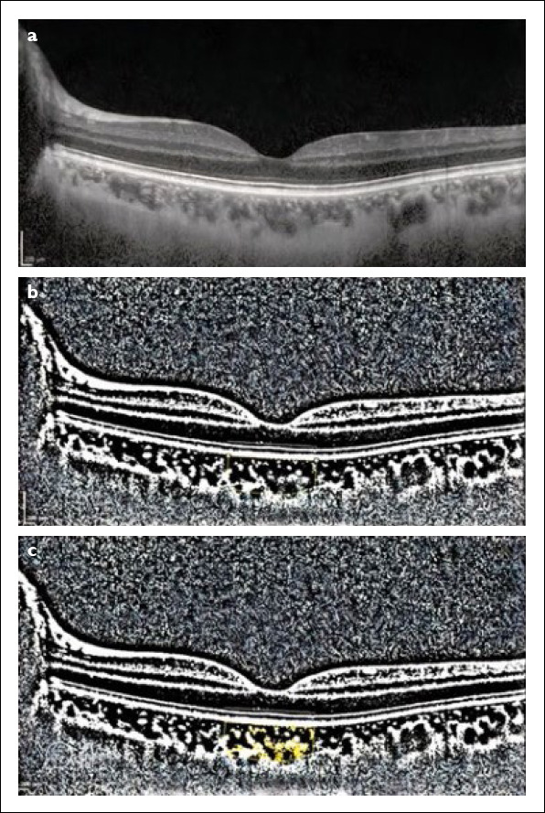
Methods: 46 epilepsy patients and 50 sex- and age-matched control patients were analyzed in the study. Patients' epilepsy types were recorded. The central macular thickness (CMT), retinal nerve fiber layer (RNFL), ganglion cell layer (GCL), and choroidal thickness (CT) were investigated through the Spectralis-OCT device (SD-OCT). Image-J program was used to calculate the total choroidal area (TCA), the luminal area (LA), stromal area (SA), and the choroidal vascularity index (CVI).
Results: CMT, TCA, LA, and SA outcomes were substantially reduced in epilepsy patients than in healthy controls. There was no significant difference between CT, RNFL, GCL, CVI results. There were no statistically significant differences between patients with partial and generalized epilepsy (p>0.05 for each).
Conclusion: According to the results of our study, epilepsy disease has effects on the posterior segment of the eye. To the best of our knowledge, our study is the first to evaluate CVI in patients with epilepsy and the epilepsy subgroups.


 分享
分享

 求助内容:
求助内容: 应助结果提醒方式:
 扫码关注我们
