Eric L. Wu MD, Atur Patel MD, Mary C. McGunigal MD, Stephanie Y. Johng MD, Armin Mortazavi MD, Ann K. Jay MD, Peter H. Ahn MD, Amjad N. Anaizi MD, Timothy R. DeKlotz MD
{"title":"Characterization of cerebral radiation necrosis following the treatment of sinonasal malignancies","authors":"Eric L. Wu MD, Atur Patel MD, Mary C. McGunigal MD, Stephanie Y. Johng MD, Armin Mortazavi MD, Ann K. Jay MD, Peter H. Ahn MD, Amjad N. Anaizi MD, Timothy R. DeKlotz MD","doi":"10.1002/lio2.1200","DOIUrl":null,"url":null,"abstract":"<div>\n \n \n <section>\n \n <h3> Objectives</h3>\n \n <p>Our study aims to determine the incidence and potential risk factors for cerebral radiation necrosis (CRN) following treatment of sinonasal malignancies.</p>\n </section>\n \n <section>\n \n <h3> Methods</h3>\n \n <p>One hundred thirty-two patients diagnosed with sinonasal malignancies over an 18-year period were identified at two institutions. Forty-six patients meeting inclusion criteria and treated with radiation therapy were included for analysis. Demographic and clinical-pathologic characteristics were collected and reviewed. Post-treatment magnetic resonance imaging (MRI) at least 1 year following treatment was reviewed to determine presence or absence of CRN.</p>\n </section>\n \n <section>\n \n <h3> Results</h3>\n \n <p>CRN was identified on MRI in 8 of 46 patients (17.4%) following radiation treatment. Patients with a history of reirradiation were more likely to develop CRN (50% vs. 10.5%, <i>p</i> < .05). The BEDs of radiation were also higher in CRN patients compared to non-CRN patients, but this difference was not significant (<i>p</i> > .05). CRN patients had a higher proportion of tumors with skull base involvement than non-CRN patients (100% vs. 57.9%, <i>p</i> = .037). Demographics, comorbidities, pathology, primary tumor subsite, chemotherapy use, and stage of disease demonstrated no significant increase in risk of CRN.</p>\n </section>\n \n <section>\n \n <h3> Conclusions</h3>\n \n <p>Reirradiation and tumor skull base involvement were significant risk factors associated with CRN. Higher average total prescribed and BEDs of radiation were seen in the CRN groups, but these differences were not statistically significant. Gender, comorbidities, tumor subsite, tumor location, and treatment type were not significantly different between groups.</p>\n </section>\n \n <section>\n \n <h3> Level of evidence</h3>\n \n <p>Level 3.</p>\n </section>\n </div>","PeriodicalId":48529,"journal":{"name":"Laryngoscope Investigative Otolaryngology","volume":"9 2","pages":""},"PeriodicalIF":1.7000,"publicationDate":"2024-03-23","publicationTypes":"Journal Article","fieldsOfStudy":null,"isOpenAccess":false,"openAccessPdf":"https://onlinelibrary.wiley.com/doi/epdf/10.1002/lio2.1200","citationCount":"0","resultStr":null,"platform":"Semanticscholar","paperid":null,"PeriodicalName":"Laryngoscope Investigative Otolaryngology","FirstCategoryId":"3","ListUrlMain":"https://onlinelibrary.wiley.com/doi/10.1002/lio2.1200","RegionNum":4,"RegionCategory":"医学","ArticlePicture":[],"TitleCN":null,"AbstractTextCN":null,"PMCID":null,"EPubDate":"","PubModel":"","JCR":"Q2","JCRName":"OTORHINOLARYNGOLOGY","Score":null,"Total":0}
引用次数: 0
Abstract
Objectives
Our study aims to determine the incidence and potential risk factors for cerebral radiation necrosis (CRN) following treatment of sinonasal malignancies.
Methods
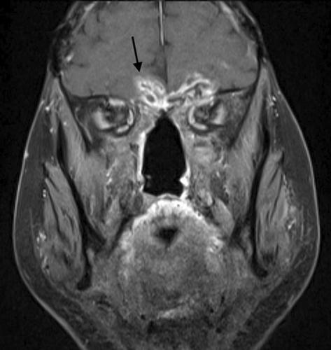
One hundred thirty-two patients diagnosed with sinonasal malignancies over an 18-year period were identified at two institutions. Forty-six patients meeting inclusion criteria and treated with radiation therapy were included for analysis. Demographic and clinical-pathologic characteristics were collected and reviewed. Post-treatment magnetic resonance imaging (MRI) at least 1 year following treatment was reviewed to determine presence or absence of CRN.
Results
CRN was identified on MRI in 8 of 46 patients (17.4%) following radiation treatment. Patients with a history of reirradiation were more likely to develop CRN (50% vs. 10.5%, p < .05). The BEDs of radiation were also higher in CRN patients compared to non-CRN patients, but this difference was not significant (p > .05). CRN patients had a higher proportion of tumors with skull base involvement than non-CRN patients (100% vs. 57.9%, p = .037). Demographics, comorbidities, pathology, primary tumor subsite, chemotherapy use, and stage of disease demonstrated no significant increase in risk of CRN.
Conclusions
Reirradiation and tumor skull base involvement were significant risk factors associated with CRN. Higher average total prescribed and BEDs of radiation were seen in the CRN groups, but these differences were not statistically significant. Gender, comorbidities, tumor subsite, tumor location, and treatment type were not significantly different between groups.


 分享
分享
 求助内容:
求助内容: 应助结果提醒方式:
 扫码关注我们
