Artificial intelligence algorithm accurately assesses oestrogen receptor immunohistochemistry in metastatic breast cancer cytology specimens: A pilot study
Brenna C. Li, Scott Hammond, Anil V. Parwani, Rulong Shen
{"title":"Artificial intelligence algorithm accurately assesses oestrogen receptor immunohistochemistry in metastatic breast cancer cytology specimens: A pilot study","authors":"Brenna C. Li, Scott Hammond, Anil V. Parwani, Rulong Shen","doi":"10.1111/cyt.13373","DOIUrl":null,"url":null,"abstract":"<div>\n \n \n <section>\n \n <h3> Objective</h3>\n \n <p>The Visiopharm artificial intelligence (AI) algorithm for oestrogen receptor (ER) immunohistochemistry (IHC) in whole slide images (WSIs) has been successfully validated in surgical pathology. This study aimed to assess its efficacy in cytology specimens.</p>\n </section>\n \n <section>\n \n <h3> Methods</h3>\n \n <p>The study cohort comprised 105 consecutive cytology specimens with metastatic breast carcinoma. ER IHC WSIs were seamlessly integrated into the Visiopharm platform from the Image Management System (IMS) during our routine digital workflow, and an AI algorithm was employed for analysis. ER AI scores were compared with pathologists' manual consensus scores. Optimization steps were implemented and evaluated to reduce discordance.</p>\n </section>\n \n <section>\n \n <h3> Results</h3>\n \n <p>The overall concordance between pathologists' scores and AI scores was excellent (99/105, 94.3%). Six cases exhibited discordant results, including two false-negative (FN) cases due to abundant histiocytes incorrectly counted as negatively stained tumour cells by AI, two FN cases owing to weak staining, and two false-positive (FP) cases where pigmented macrophages were erroneously counted as positively stained tumour cells by AI. The Pearson correlation coefficient of ER-positive percentages between pathologists' and AI scores was 0.8483. Optimization steps, such as lowering the cut-off threshold and additional training using higher input magnification, significantly improved accuracy.</p>\n </section>\n \n <section>\n \n <h3> Conclusions</h3>\n \n <p>The automated ER AI algorithm demonstrated excellent concordance with pathologists' assessments and accurately differentiated ER-positive from ER-negative metastatic breast carcinoma cytology cases. However, precision in identifying tumour cells in cytology specimens requires further enhancement.</p>\n </section>\n </div>","PeriodicalId":55187,"journal":{"name":"Cytopathology","volume":"35 4","pages":"464-472"},"PeriodicalIF":1.1000,"publicationDate":"2024-03-22","publicationTypes":"Journal Article","fieldsOfStudy":null,"isOpenAccess":false,"openAccessPdf":"","citationCount":"0","resultStr":null,"platform":"Semanticscholar","paperid":null,"PeriodicalName":"Cytopathology","FirstCategoryId":"3","ListUrlMain":"https://onlinelibrary.wiley.com/doi/10.1111/cyt.13373","RegionNum":4,"RegionCategory":"医学","ArticlePicture":[],"TitleCN":null,"AbstractTextCN":null,"PMCID":null,"EPubDate":"","PubModel":"","JCR":"Q4","JCRName":"CELL BIOLOGY","Score":null,"Total":0}
引用次数: 0
Abstract
Objective
The Visiopharm artificial intelligence (AI) algorithm for oestrogen receptor (ER) immunohistochemistry (IHC) in whole slide images (WSIs) has been successfully validated in surgical pathology. This study aimed to assess its efficacy in cytology specimens.
Methods
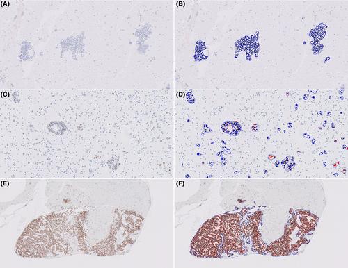
The study cohort comprised 105 consecutive cytology specimens with metastatic breast carcinoma. ER IHC WSIs were seamlessly integrated into the Visiopharm platform from the Image Management System (IMS) during our routine digital workflow, and an AI algorithm was employed for analysis. ER AI scores were compared with pathologists' manual consensus scores. Optimization steps were implemented and evaluated to reduce discordance.
Results
The overall concordance between pathologists' scores and AI scores was excellent (99/105, 94.3%). Six cases exhibited discordant results, including two false-negative (FN) cases due to abundant histiocytes incorrectly counted as negatively stained tumour cells by AI, two FN cases owing to weak staining, and two false-positive (FP) cases where pigmented macrophages were erroneously counted as positively stained tumour cells by AI. The Pearson correlation coefficient of ER-positive percentages between pathologists' and AI scores was 0.8483. Optimization steps, such as lowering the cut-off threshold and additional training using higher input magnification, significantly improved accuracy.
Conclusions
The automated ER AI algorithm demonstrated excellent concordance with pathologists' assessments and accurately differentiated ER-positive from ER-negative metastatic breast carcinoma cytology cases. However, precision in identifying tumour cells in cytology specimens requires further enhancement.
期刊介绍:
The aim of Cytopathology is to publish articles relating to those aspects of cytology which will increase our knowledge and understanding of the aetiology, diagnosis and management of human disease. It contains original articles and critical reviews on all aspects of clinical cytology in its broadest sense, including: gynaecological and non-gynaecological cytology; fine needle aspiration and screening strategy.
Cytopathology welcomes papers and articles on: ultrastructural, histochemical and immunocytochemical studies of the cell; quantitative cytology and DNA hybridization as applied to cytological material.


 分享
分享
 求助内容:
求助内容: 应助结果提醒方式:
 扫码关注我们
