{"title":"Accessory lingual mental foramen: A case report of a rare anatomic variation.","authors":"Arjun Kumar Tallada, Junaid Ahmed, Nanditha Sujir, Nandita Shenoy, Shubham M Pawar, Archana Muralidharan, Sanjay Mallya","doi":"10.1007/s11282-024-00747-5","DOIUrl":null,"url":null,"abstract":"<p><strong>Introduction: </strong>The mandibular nerve and the mental foramen have occasionally shown variations in its anatomy. This report aims to present a case of lingual mental foramen recognised on three-dimensional cone beam computed tomographic imaging (CBCT).</p><p><strong>Case report: </strong>Routine Orthopantomogram (OPG) and CBCT images were evaluated to assess the status of impact third molars in a 31-year-old female who had visited the dental clinics in our institution. The OPG image failed to reveal any anatomic variation in the position of the mental foramen. On tracing the course of the mandibular canal in CBCT images, two foramina were traced at the region of premolar. One opened towards the buccal cortical plate at the normal position of the mental foramen and an accessory lingual mental foramen had an opening on the lingual cortical bone at the same level as the mental foramen.</p><p><strong>Conclusion: </strong>Understanding variations of the mental foramen is extremely essential in dentistry to carry out successful anaesthetic or surgical interventions and to avoid complications such as nerve damage or excessive bleeding.</p>","PeriodicalId":56103,"journal":{"name":"Oral Radiology","volume":" ","pages":"410-414"},"PeriodicalIF":1.7000,"publicationDate":"2024-07-01","publicationTypes":"Journal Article","fieldsOfStudy":null,"isOpenAccess":false,"openAccessPdf":"","citationCount":"0","resultStr":null,"platform":"Semanticscholar","paperid":null,"PeriodicalName":"Oral Radiology","FirstCategoryId":"3","ListUrlMain":"https://doi.org/10.1007/s11282-024-00747-5","RegionNum":3,"RegionCategory":"医学","ArticlePicture":[],"TitleCN":null,"AbstractTextCN":null,"PMCID":null,"EPubDate":"2024/3/25 0:00:00","PubModel":"Epub","JCR":"Q3","JCRName":"DENTISTRY, ORAL SURGERY & MEDICINE","Score":null,"Total":0}
引用次数: 0
Abstract
Introduction: The mandibular nerve and the mental foramen have occasionally shown variations in its anatomy. This report aims to present a case of lingual mental foramen recognised on three-dimensional cone beam computed tomographic imaging (CBCT).
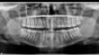
Case report: Routine Orthopantomogram (OPG) and CBCT images were evaluated to assess the status of impact third molars in a 31-year-old female who had visited the dental clinics in our institution. The OPG image failed to reveal any anatomic variation in the position of the mental foramen. On tracing the course of the mandibular canal in CBCT images, two foramina were traced at the region of premolar. One opened towards the buccal cortical plate at the normal position of the mental foramen and an accessory lingual mental foramen had an opening on the lingual cortical bone at the same level as the mental foramen.
Conclusion: Understanding variations of the mental foramen is extremely essential in dentistry to carry out successful anaesthetic or surgical interventions and to avoid complications such as nerve damage or excessive bleeding.
期刊介绍:
As the official English-language journal of the Japanese Society for Oral and Maxillofacial Radiology and the Asian Academy of Oral and Maxillofacial Radiology, Oral Radiology is intended to be a forum for international collaboration in head and neck diagnostic imaging and all related fields. Oral Radiology features cutting-edge research papers, review articles, case reports, and technical notes from both the clinical and experimental fields. As membership in the Society is not a prerequisite, contributions are welcome from researchers and clinicians worldwide.


 分享
分享
 求助内容:
求助内容: 应助结果提醒方式:
 扫码关注我们
