Combined positron emission tomography-guided modified black phosphorus nanosheet-based photothermal therapy and anti programmed cell death protein ligand 1 therapy
{"title":"Combined positron emission tomography-guided modified black phosphorus nanosheet-based photothermal therapy and anti programmed cell death protein ligand 1 therapy","authors":"Xiaona Sun, Siqi Zhang, Shuo Jiang, Jieting Shen, Yuxuan Wu, Hailong Zhang, Ming-Rong Zhang, Rui Wang, Kuan Hu","doi":"10.1002/ird3.69","DOIUrl":null,"url":null,"abstract":"<div>\n \n \n <section>\n \n <h3> Background</h3>\n \n <p>Patients with <i>cold</i> tumors gain limited benefits from immune checkpoint blockade (ICB) therapy owing to low programmed cell death protein ligand 1 (PD-L1) expression and minimal immune cell infiltration. Mild photothermal therapy (PTT) using black phosphorus nanosheets (BPNSs) is a promising approach to enhance the efficacy of ICB therapy. However, to ensure that BPNS-based PTT-enhanced ICB therapy is clinically adaptable, a noninvasive, bedside-accessible imaging tool capable of monitoring the status of PD-L1 is imperative. We demonstrated that positron emission tomography (PET) using [<sup>64</sup>Cu]HKP2202 precisely delineated PD-L1 expression in tumors receiving PTT.</p>\n </section>\n \n <section>\n \n <h3> Methods</h3>\n \n <p>BPNSs were modified with polyethylene glycol to prepare BPNS@PEG, which were then characterized. MC38 cells and tumor allografts were treated with BPNS@PEG followed by 808 nm near-infrared light exposure. PET using [<sup>64</sup>Cu]HKP2202 was performed to monitor PD-L1 expression in vivo. We also evaluated whether the efficacy of ICB therapy improved after delivering BPNS@PEG-based PTT.</p>\n </section>\n \n <section>\n \n <h3> Results</h3>\n \n <p>BPNS@PEG had a well-defined lamellar structure with clear edges and an average size of 150 nm. PET and Western blotting assays indicated that PD-L1 expression was upregulated after BPNS@PEG and NIR-light treatment. Notably, the antitumor effect of anti PD-L1 therapy was enhanced in mice treated with BPNS@PEG-based PTT.</p>\n </section>\n \n <section>\n \n <h3> Conclusions</h3>\n \n <p>BPNS@PEG had the capacity to convert <i>cold</i> tumors into <i>hot</i> tumors to facilitate the efficacy of ICB therapy. Importantly, the complementary diagnostic PET radiotracer targeting PD-L1 allowed real-time monitoring of PD-L1 expression in the tumor microenvironment to guide ICB administration, holding great potential to achieve efficient and precise tumor immunotherapy.</p>\n </section>\n </div>","PeriodicalId":73508,"journal":{"name":"iRadiology","volume":"2 2","pages":"103-112"},"PeriodicalIF":0.0000,"publicationDate":"2024-03-13","publicationTypes":"Journal Article","fieldsOfStudy":null,"isOpenAccess":false,"openAccessPdf":"https://onlinelibrary.wiley.com/doi/epdf/10.1002/ird3.69","citationCount":"0","resultStr":null,"platform":"Semanticscholar","paperid":null,"PeriodicalName":"iRadiology","FirstCategoryId":"1085","ListUrlMain":"https://onlinelibrary.wiley.com/doi/10.1002/ird3.69","RegionNum":0,"RegionCategory":null,"ArticlePicture":[],"TitleCN":null,"AbstractTextCN":null,"PMCID":null,"EPubDate":"","PubModel":"","JCR":"","JCRName":"","Score":null,"Total":0}
引用次数: 0
Abstract
Background
Patients with cold tumors gain limited benefits from immune checkpoint blockade (ICB) therapy owing to low programmed cell death protein ligand 1 (PD-L1) expression and minimal immune cell infiltration. Mild photothermal therapy (PTT) using black phosphorus nanosheets (BPNSs) is a promising approach to enhance the efficacy of ICB therapy. However, to ensure that BPNS-based PTT-enhanced ICB therapy is clinically adaptable, a noninvasive, bedside-accessible imaging tool capable of monitoring the status of PD-L1 is imperative. We demonstrated that positron emission tomography (PET) using [64Cu]HKP2202 precisely delineated PD-L1 expression in tumors receiving PTT.
Methods
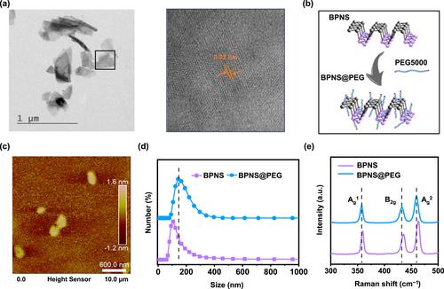
BPNSs were modified with polyethylene glycol to prepare BPNS@PEG, which were then characterized. MC38 cells and tumor allografts were treated with BPNS@PEG followed by 808 nm near-infrared light exposure. PET using [64Cu]HKP2202 was performed to monitor PD-L1 expression in vivo. We also evaluated whether the efficacy of ICB therapy improved after delivering BPNS@PEG-based PTT.
Results
BPNS@PEG had a well-defined lamellar structure with clear edges and an average size of 150 nm. PET and Western blotting assays indicated that PD-L1 expression was upregulated after BPNS@PEG and NIR-light treatment. Notably, the antitumor effect of anti PD-L1 therapy was enhanced in mice treated with BPNS@PEG-based PTT.
Conclusions
BPNS@PEG had the capacity to convert cold tumors into hot tumors to facilitate the efficacy of ICB therapy. Importantly, the complementary diagnostic PET radiotracer targeting PD-L1 allowed real-time monitoring of PD-L1 expression in the tumor microenvironment to guide ICB administration, holding great potential to achieve efficient and precise tumor immunotherapy.


 分享
分享

 求助内容:
求助内容: 应助结果提醒方式:
 扫码关注我们
