A Low-Risk HPV-Associated Well-Differentiated Squamous Cell Carcinoma of the Cervix with Low-Grade Squamous Intraepithelial Lesion Morphology: Clinical and Pathologic Diagnostic Difficulties and Review of the Literature.
Deniz Ates, Esra Nur Sahin, Kübra Katipoglu, Alp Usubutun
{"title":"A Low-Risk HPV-Associated Well-Differentiated Squamous Cell Carcinoma of the Cervix with Low-Grade Squamous Intraepithelial Lesion Morphology: Clinical and Pathologic Diagnostic Difficulties and Review of the Literature.","authors":"Deniz Ates, Esra Nur Sahin, Kübra Katipoglu, Alp Usubutun","doi":"10.5146/tjpath.2024.13189","DOIUrl":null,"url":null,"abstract":"<p><p>Approximately 95% of cervical squamous cell carcinomas are associated with high-risk HPV, with a small number of HPV-independent tumors. However, low-risk HPV types have also been detected in rare cervical squamous cell carcinomas. Low-grade squamous intraepithelial lesion-related changes are a rare morphologic finding in cervical squamous cell carcinoma. We present the case of a 30-yr-old woman who presented with pelvic pain and foul-smelling vaginal discharge showing an exophytic lesion protruding from the cervix. Repeated superficial biopsies showed a low-grade squamous intraepithelial lesion (LSIL) characterized by binucleation and koilocytosis. Chromogenic in-situ hybridization revealed the presence of HPV6/11. The absence of high-risk HPV was confirmed by PCR. After following the patient for nine months without intervention, type III hysterectomy and bilateral pelvic paraaortic lymphadenectomy were performed. Microscopic examination showed well-differentiated squamous cell carcinoma with solid epithelial islands and extensive eosinophilic cytoplasm without pleomorphism. HPV 6 and 11 were also detected with chromogenic in-situ hybridization. Neoplasm invaded the full-thickness of the cervical wall and infiltrated the vagina, parametrium, the proximal ureter and bladder. The patient who received chemoradiotherapy is disease-free at 36 months follow-up. Low-risk HPV-related well-differentiated invasive squamous lesions exist, and such lesions could be a diagnostic pitfall for gynecologists and pathologists; in these cases, radiologic-pathologic correlation and radiologic guided biopsy are mandatory.</p>","PeriodicalId":45415,"journal":{"name":"Turkish Journal of Pathology","volume":" ","pages":"196-201"},"PeriodicalIF":2.1000,"publicationDate":"2024-01-01","publicationTypes":"Journal Article","fieldsOfStudy":null,"isOpenAccess":false,"openAccessPdf":"https://www.ncbi.nlm.nih.gov/pmc/articles/PMC11391893/pdf/","citationCount":"0","resultStr":null,"platform":"Semanticscholar","paperid":null,"PeriodicalName":"Turkish Journal of Pathology","FirstCategoryId":"1085","ListUrlMain":"https://doi.org/10.5146/tjpath.2024.13189","RegionNum":0,"RegionCategory":null,"ArticlePicture":[],"TitleCN":null,"AbstractTextCN":null,"PMCID":null,"EPubDate":"","PubModel":"","JCR":"Q4","JCRName":"PATHOLOGY","Score":null,"Total":0}
引用次数: 0
Abstract
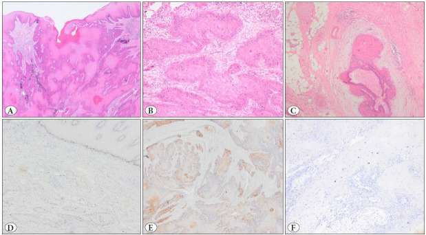
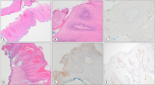
Approximately 95% of cervical squamous cell carcinomas are associated with high-risk HPV, with a small number of HPV-independent tumors. However, low-risk HPV types have also been detected in rare cervical squamous cell carcinomas. Low-grade squamous intraepithelial lesion-related changes are a rare morphologic finding in cervical squamous cell carcinoma. We present the case of a 30-yr-old woman who presented with pelvic pain and foul-smelling vaginal discharge showing an exophytic lesion protruding from the cervix. Repeated superficial biopsies showed a low-grade squamous intraepithelial lesion (LSIL) characterized by binucleation and koilocytosis. Chromogenic in-situ hybridization revealed the presence of HPV6/11. The absence of high-risk HPV was confirmed by PCR. After following the patient for nine months without intervention, type III hysterectomy and bilateral pelvic paraaortic lymphadenectomy were performed. Microscopic examination showed well-differentiated squamous cell carcinoma with solid epithelial islands and extensive eosinophilic cytoplasm without pleomorphism. HPV 6 and 11 were also detected with chromogenic in-situ hybridization. Neoplasm invaded the full-thickness of the cervical wall and infiltrated the vagina, parametrium, the proximal ureter and bladder. The patient who received chemoradiotherapy is disease-free at 36 months follow-up. Low-risk HPV-related well-differentiated invasive squamous lesions exist, and such lesions could be a diagnostic pitfall for gynecologists and pathologists; in these cases, radiologic-pathologic correlation and radiologic guided biopsy are mandatory.


 分享
分享
 求助内容:
求助内容: 应助结果提醒方式:
 扫码关注我们
