Carolina Alonso Amorim, Marília F Marceliano-Alves, Isabelle Louise Gomes, José C Provenzano, Flávio R F Alves
{"title":"Natural canal deviation and dentin thickness of mesial root canals of mandibular first molars assessed by microcomputed tomography.","authors":"Carolina Alonso Amorim, Marília F Marceliano-Alves, Isabelle Louise Gomes, José C Provenzano, Flávio R F Alves","doi":"10.1590/0103-6440202405648","DOIUrl":null,"url":null,"abstract":"<p><p>The aim of this study was to assess the centralization and dentin thickness of mesial root canals of the first mandibular molars by microcomputed tomography (micro-CT). Material and methods: Ninety-nine mandibular molars of Vertucci's type IV canals were scanned by micro-CT. The mesiodistal deviation and centroid were assessed, in both mesiobuccal (MB) and mesiolingual (ML) canals, for the apical 4mm and the full canal length. Results: The dentin thickness was similar for both MB and ML canals. The narrowest thickness was in the distal wall of an MB canal (0.07mm), while the widest was found in the mesial wall of an MB canal (2.46mm). In centroid analysis, both the MB and ML canals exhibited deviations when compared to the root centroid, along the full canal length and the apical 4mm. For the MB canal, the mean deviation was 0.83mm (0.02 mm-2.30 mm) for the full canal and 0.18mm (0.01 mm-1.01 mm) for apical 4mm. Similarly, for the ML canal, the mean deviation measured 0.83 mm (0.05mm-3.99mm) for the full canal and 0.21 mm (0.01mm-1.01mm) for the apical 4 mm. Overall, deviations were observed towards the mesial of the roots, with 69% for MB and 57% for ML canals for the full canal, and 51% for MB canals within the 4 mm. The exception was the ML canal, which exhibited a higher deviation towards distal in the apical 4mm, accounting for 52% of cases. The dentin thickness was consistent between the mesial canals of mandibular molars. However, there is no centrality of mesial canals in their roots, with frequent deviation to mesial.</p>","PeriodicalId":101363,"journal":{"name":"Brazilian dental journal","volume":"35 ","pages":"e245648"},"PeriodicalIF":0.0000,"publicationDate":"2024-03-22","publicationTypes":"Journal Article","fieldsOfStudy":null,"isOpenAccess":false,"openAccessPdf":"https://www.ncbi.nlm.nih.gov/pmc/articles/PMC10976306/pdf/","citationCount":"0","resultStr":null,"platform":"Semanticscholar","paperid":null,"PeriodicalName":"Brazilian dental journal","FirstCategoryId":"1085","ListUrlMain":"https://doi.org/10.1590/0103-6440202405648","RegionNum":0,"RegionCategory":null,"ArticlePicture":[],"TitleCN":null,"AbstractTextCN":null,"PMCID":null,"EPubDate":"2024/1/1 0:00:00","PubModel":"eCollection","JCR":"","JCRName":"","Score":null,"Total":0}
引用次数: 0
Abstract
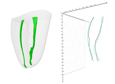
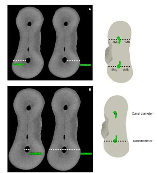
The aim of this study was to assess the centralization and dentin thickness of mesial root canals of the first mandibular molars by microcomputed tomography (micro-CT). Material and methods: Ninety-nine mandibular molars of Vertucci's type IV canals were scanned by micro-CT. The mesiodistal deviation and centroid were assessed, in both mesiobuccal (MB) and mesiolingual (ML) canals, for the apical 4mm and the full canal length. Results: The dentin thickness was similar for both MB and ML canals. The narrowest thickness was in the distal wall of an MB canal (0.07mm), while the widest was found in the mesial wall of an MB canal (2.46mm). In centroid analysis, both the MB and ML canals exhibited deviations when compared to the root centroid, along the full canal length and the apical 4mm. For the MB canal, the mean deviation was 0.83mm (0.02 mm-2.30 mm) for the full canal and 0.18mm (0.01 mm-1.01 mm) for apical 4mm. Similarly, for the ML canal, the mean deviation measured 0.83 mm (0.05mm-3.99mm) for the full canal and 0.21 mm (0.01mm-1.01mm) for the apical 4 mm. Overall, deviations were observed towards the mesial of the roots, with 69% for MB and 57% for ML canals for the full canal, and 51% for MB canals within the 4 mm. The exception was the ML canal, which exhibited a higher deviation towards distal in the apical 4mm, accounting for 52% of cases. The dentin thickness was consistent between the mesial canals of mandibular molars. However, there is no centrality of mesial canals in their roots, with frequent deviation to mesial.


 分享
分享

 求助内容:
求助内容: 应助结果提醒方式:
 扫码关注我们
