Ming Wang, Binyuan Xu, Yangmei Xie, Ge Yao, Yinghui Chen
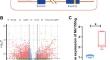
{"title":"Mir155hg Accelerates Hippocampal Neuron Injury in Convulsive Status Epilepticus by Inhibiting Microglial Phagocytosis","authors":"Ming Wang, Binyuan Xu, Yangmei Xie, Ge Yao, Yinghui Chen","doi":"10.1007/s11064-024-04131-x","DOIUrl":null,"url":null,"abstract":"<div><p>Convulsive status epilepticus (CSE) is a common critical neurological condition that can lead to irreversible hippocampal neuron damage and cognitive dysfunction. Multiple studies have demonstrated the critical roles that long non-coding RNA Mir155hg plays in a variety of diseases. However, less is known about the function and mechanism of Mir155hg in CSE. Here we investigate and elucidate the mechanism underlying the contribution of Mir155hg to CSE-induced hippocampal neuron injury. By applying high-throughput sequencing, we examined the expression of differentially expressed genes in normal and CSE rats. Subsequent RT-qPCR enabled us to measure the level of Mir155hg in rat hippocampal tissue. Targeted knockdown of Mir155hg was achieved by the AAV9 virus. Additionally, we utilized HE and Tunel staining to evaluate neuronal injury. Immunofluorescence (IF), Golgi staining, and brain path clamping were also used to detect the synaptic plasticity of hippocampal neurons. Finally, through IF staining and Sholl analysis, we assessed the degree of microglial phagocytic function. It was found that the expression of Mir155hg was elevated in CSE rats. HE and Tunel staining results showed that Mir155hg knockdown suppressed the hippocampal neuron loss and apoptosis followed CSE. IF, Golgi staining and brain path clamp data found that Mir155hg knockdown enhanced neuronal synaptic plasticity. The results from IF staining and Sholl analysis showed that Mir155hg knockdown enhanced microglial phagocytosis. Our findings suggest that Mir155hg promotes CSE-induced hippocampal neuron injury by inhibiting microglial phagocytosis.</p></div>","PeriodicalId":719,"journal":{"name":"Neurochemical Research","volume":"49 7","pages":"1782 - 1793"},"PeriodicalIF":3.8000,"publicationDate":"2024-03-30","publicationTypes":"Journal Article","fieldsOfStudy":null,"isOpenAccess":false,"openAccessPdf":"","citationCount":"0","resultStr":null,"platform":"Semanticscholar","paperid":null,"PeriodicalName":"Neurochemical Research","FirstCategoryId":"3","ListUrlMain":"https://link.springer.com/article/10.1007/s11064-024-04131-x","RegionNum":3,"RegionCategory":"医学","ArticlePicture":[],"TitleCN":null,"AbstractTextCN":null,"PMCID":null,"EPubDate":"","PubModel":"","JCR":"Q2","JCRName":"BIOCHEMISTRY & MOLECULAR BIOLOGY","Score":null,"Total":0}
引用次数: 0
Abstract
Convulsive status epilepticus (CSE) is a common critical neurological condition that can lead to irreversible hippocampal neuron damage and cognitive dysfunction. Multiple studies have demonstrated the critical roles that long non-coding RNA Mir155hg plays in a variety of diseases. However, less is known about the function and mechanism of Mir155hg in CSE. Here we investigate and elucidate the mechanism underlying the contribution of Mir155hg to CSE-induced hippocampal neuron injury. By applying high-throughput sequencing, we examined the expression of differentially expressed genes in normal and CSE rats. Subsequent RT-qPCR enabled us to measure the level of Mir155hg in rat hippocampal tissue. Targeted knockdown of Mir155hg was achieved by the AAV9 virus. Additionally, we utilized HE and Tunel staining to evaluate neuronal injury. Immunofluorescence (IF), Golgi staining, and brain path clamping were also used to detect the synaptic plasticity of hippocampal neurons. Finally, through IF staining and Sholl analysis, we assessed the degree of microglial phagocytic function. It was found that the expression of Mir155hg was elevated in CSE rats. HE and Tunel staining results showed that Mir155hg knockdown suppressed the hippocampal neuron loss and apoptosis followed CSE. IF, Golgi staining and brain path clamp data found that Mir155hg knockdown enhanced neuronal synaptic plasticity. The results from IF staining and Sholl analysis showed that Mir155hg knockdown enhanced microglial phagocytosis. Our findings suggest that Mir155hg promotes CSE-induced hippocampal neuron injury by inhibiting microglial phagocytosis.
期刊介绍:
Neurochemical Research is devoted to the rapid publication of studies that use neurochemical methodology in research on nervous system structure and function. The journal publishes original reports of experimental and clinical research results, perceptive reviews of significant problem areas in the neurosciences, brief comments of a methodological or interpretive nature, and research summaries conducted by leading scientists whose works are not readily available in English.


 分享
分享
 求助内容:
求助内容: 应助结果提醒方式:
 扫码关注我们
