Knowledge of the physical effects of pulsed dye laser (PDL) treatment of psoriatic lesions is essential in unraveling the remedial mechanisms of this treatment and hence also in maximizing in its disease-modifying potential. Therefore, the main objective of this study was to provide estimates of these physical effects (for laser wavelengths of 585 and 595 nm), with the aim of identifying pathogenic processes that may be affected by these conditions.
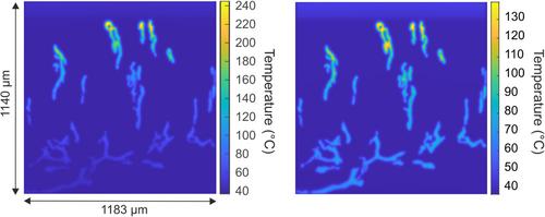
We modeled the laser light propagation and subsequent photothermal heating by numerically solving the transient diffusion and heat equations simultaneously. To this end, we used the finite element method in conjunction with an image-derived psoriatic lesion morphology (which was defined by segmenting blood vessels from a confocal microscopy image of a fluorescently labeled section of a 3 mm punch biopsy of a psoriatic lesion). The resulting predictions of the generated temperature field within the lesion were then used to assess the possibility of stalling or arresting some suspected pathogenic processes.
According to our results, it is conceivable that perivascular nerves are thermally denatured, as almost all locations that reach 60°C were found to be within 18 µm (at 585 nm) and 11 µm (at 595 nm) of a blood vessel wall. Furthermore, activation of TRPV1 and TRPV2 channels in perivascular neuronal and immune cells is highly likely, since a critical temperature of 43°C is generated at locations within up to 350 µm of a vessel wall (at both wavelengths) and sustained for up to 700 ms (at 585 nm) and 40 ms (at 595 nm), while a critical temperature of 52°C is reached by locations within 80 µm (at 585 nm) and 30 µm (at 595 nm) of a vessel wall and sustained for up to 100 ms (at 585 nm) and 30 ms (at 595 nm). Finally, we found that the blood vessel coagulation-inducing temperature of 70°C is sustained in the vascular epithelium for up to 19 and 5 ms at 585 and 595 nm, respectively, rendering partial or total loss of vascular functionality a distinct possibility.
The presented approach constitutes a useful tool to provide realistic estimates of the photothermal effects of PDL treatment of psoriatic plaques (as well as other selective photothermolysis-based treatments), yielding information that is essential in guiding future experimental studies toward unraveling the remedial mechanisms of these treatments.


 分享
分享
 求助内容:
求助内容: 应助结果提醒方式:
 扫码关注我们
