Vitamin D3 reduces the symptoms of ovarian hyperstimulation syndrome in mice and inhibits the release of granulosa cell angiogenic factor through pentraxin 3
Minping Zhang, Li Chen, Qunping Xu, Xiaohua Yang, Xiang Liu, Luanmei Liu
{"title":"Vitamin D3 reduces the symptoms of ovarian hyperstimulation syndrome in mice and inhibits the release of granulosa cell angiogenic factor through pentraxin 3","authors":"Minping Zhang, Li Chen, Qunping Xu, Xiaohua Yang, Xiang Liu, Luanmei Liu","doi":"10.1007/s11626-024-00898-z","DOIUrl":null,"url":null,"abstract":"<p>It has been reported that the effective inhibition of vascular endothelial growth factor (VEGF) can prevent the progression of ovarian hyperstimulation syndrome (OHSS). The present study aimed to investigate the mechanism underlying the effect of vitamin D<sub>3</sub> (VD3) on OHSS in mouse models and granulosa cells. The effects of VD3 administration (16 and 24 IU) on ovarian permeability were determined using Evans blue. In addition, ovarian pathology, corpus luteum count, inflammatory responses, and hormone and VEGFA levels were assessed using pathological sections and ELISA. Molecular docking predicted that pentraxin 3 (PTX3) could be a potential target of VD3, and therefore, the effects of human chorionic gonadotropin (hCG) and VD3 as well as PTX3 overexpression on the production and secretion of VEGFA in granulosa cells were also investigated using western blotting and immunofluorescence. Twenty-four IU VD3 significantly reversed the increase in ovarian weight and permeability in mice with OHSS. Additionally, VD3 diminished congestion and the number of corpus luteum in the ovaries and reduced the secretion levels of inflammatory factors and those of estrogen and progesterone. Notably, VD3 downregulated VEGFA and CD31 in ovarian tissues, while the expression levels of PTX3 varied among different groups. Furthermore, VD3 restored the hCG-induced enhanced VEGFA and PTX3 expression levels in granulosa cells, whereas PTX3 overexpression abrogated the VD3-mediated inhibition of VEGFA production and secretion. The present study demonstrated that VD3 could inhibit the release of VEGFA through PTX3, thus supporting the beneficial effects of VD3 administration on ameliorating OHSS symptoms.</p>","PeriodicalId":13340,"journal":{"name":"In Vitro Cellular & Developmental Biology. Animal","volume":"41 1","pages":""},"PeriodicalIF":1.7000,"publicationDate":"2024-04-04","publicationTypes":"Journal Article","fieldsOfStudy":null,"isOpenAccess":false,"openAccessPdf":"","citationCount":"0","resultStr":null,"platform":"Semanticscholar","paperid":null,"PeriodicalName":"In Vitro Cellular & Developmental Biology. Animal","FirstCategoryId":"99","ListUrlMain":"https://doi.org/10.1007/s11626-024-00898-z","RegionNum":4,"RegionCategory":"生物学","ArticlePicture":[],"TitleCN":null,"AbstractTextCN":null,"PMCID":null,"EPubDate":"","PubModel":"","JCR":"Q4","JCRName":"CELL BIOLOGY","Score":null,"Total":0}
引用次数: 0
Abstract
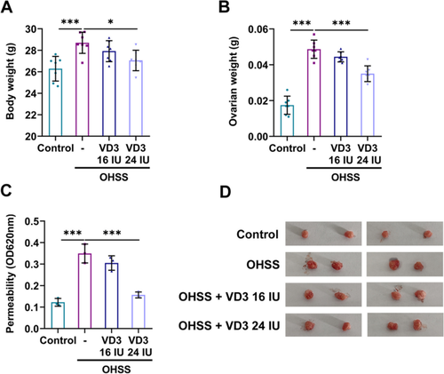
It has been reported that the effective inhibition of vascular endothelial growth factor (VEGF) can prevent the progression of ovarian hyperstimulation syndrome (OHSS). The present study aimed to investigate the mechanism underlying the effect of vitamin D3 (VD3) on OHSS in mouse models and granulosa cells. The effects of VD3 administration (16 and 24 IU) on ovarian permeability were determined using Evans blue. In addition, ovarian pathology, corpus luteum count, inflammatory responses, and hormone and VEGFA levels were assessed using pathological sections and ELISA. Molecular docking predicted that pentraxin 3 (PTX3) could be a potential target of VD3, and therefore, the effects of human chorionic gonadotropin (hCG) and VD3 as well as PTX3 overexpression on the production and secretion of VEGFA in granulosa cells were also investigated using western blotting and immunofluorescence. Twenty-four IU VD3 significantly reversed the increase in ovarian weight and permeability in mice with OHSS. Additionally, VD3 diminished congestion and the number of corpus luteum in the ovaries and reduced the secretion levels of inflammatory factors and those of estrogen and progesterone. Notably, VD3 downregulated VEGFA and CD31 in ovarian tissues, while the expression levels of PTX3 varied among different groups. Furthermore, VD3 restored the hCG-induced enhanced VEGFA and PTX3 expression levels in granulosa cells, whereas PTX3 overexpression abrogated the VD3-mediated inhibition of VEGFA production and secretion. The present study demonstrated that VD3 could inhibit the release of VEGFA through PTX3, thus supporting the beneficial effects of VD3 administration on ameliorating OHSS symptoms.
期刊介绍:
In Vitro Cellular & Developmental Biology - Animal is a journal of the Society for In Vitro Biology (SIVB). Original manuscripts reporting results of research in cellular, molecular, and developmental biology that employ or are relevant to organs, tissue, tumors, and cells in vitro will be considered for publication. Topics covered include:
Biotechnology;
Cell and Tissue Models;
Cell Growth/Differentiation/Apoptosis;
Cellular Pathology/Virology;
Cytokines/Growth Factors/Adhesion Factors;
Establishment of Cell Lines;
Signal Transduction;
Stem Cells;
Toxicology/Chemical Carcinogenesis;
Product Applications.


 分享
分享
 求助内容:
求助内容: 应助结果提醒方式:
 扫码关注我们
